 Movie
Movie Controller


+ Open data

- Basic information
| Entry | Database: PDB / ID: 3mgn | ||||||
|---|---|---|---|---|---|---|---|
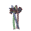
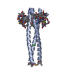
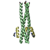
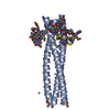
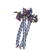
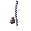
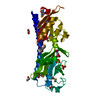
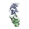
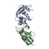
| Title | D-Peptide inhibitor PIE71 in complex with IQN17 | ||||||
 Components Components |
| ||||||
 Keywords Keywords |  VIRAL PROTEIN/VIRAL PROTEIN inhibitor / PIE71 / IQN17 / HIV / helix / coiled-coil / D-peptide inhibitor / VIRAL PROTEIN-VIRAL PROTEIN inhibitor complex VIRAL PROTEIN/VIRAL PROTEIN inhibitor / PIE71 / IQN17 / HIV / helix / coiled-coil / D-peptide inhibitor / VIRAL PROTEIN-VIRAL PROTEIN inhibitor complex | ||||||
| Function / homology | Single alpha-helices involved in coiled-coils or other helix-helix interfaces - #170 / Single alpha-helices involved in coiled-coils or other helix-helix interfaces / Up-down Bundle / Mainly Alpha / D-PEPTIDE INHIBITOR PIE71 Function and homology information Function and homology information | ||||||
| Method | X-RAY DIFFRACTION / SYNCHROTRON / MOLECULAR REPLACEMENT / Resolution: 1.4 Å | ||||||
 Authors Authors | Hill, C.P. / Whitby, F.G. / Kay, M. / Francis, N. | ||||||
 Citation Citation | Journal: J.Virol. / Year: 2010 Title: Design of a potent D-peptide HIV-1 entry inhibitor with a strong barrier to resistance. Authors: Welch, B.D. / Francis, J.N. / Redman, J.S. / Paul, S. / Weinstock, M.T. / Reeves, J.D. / Lie, Y.S. / Whitby, F.G. / Eckert, D.M. / Hill, C.P. / Root, M.J. / Kay, M.S. | ||||||
| History |
|
- Structure visualization
| Structure viewer | Molecule: MolmilJmol/JSmol |
|---|
- Downloads & links
-Download
| PDBx/mmCIF format | 3mgn.cif.gz | 92.1 KB | Display | PDBx/mmCIF format |
|---|---|---|---|---|
| PDB format | pdb3mgn.ent.gz | 81.6 KB | Display | PDB format |
| PDBx/mmJSON format | 3mgn.json.gz | Tree view | PDBx/mmJSON format | |
| Others |  Other downloads Other downloads |
-Validation report
| Arichive directory | https://data.pdbj.org/pub/pdb/validation_reports/mg/3mgnftp://data.pdbj.org/pub/pdb/validation_reports/mg/3mgn | HTTPS FTP |
|---|
-Related structure data


-Links
 PDBj
PDBj
- Assembly
| Deposited unit | 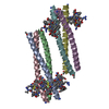
| ||||||||
|---|---|---|---|---|---|---|---|---|---|
| 1 | 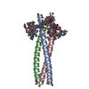
| ||||||||
| 2 | 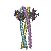
| ||||||||
| Unit cell |
|
-Components
| #1: Protein/peptide | Mass: 5466.574 Da / Num. of mol.: 6 / Source method: obtained synthetically #2: Protein/peptide |   Type: Cyclic peptide / Class: Inhibitor / Mass: 1874.236 Da / Num. of mol.: 6 / Source method: obtained synthetically / References: D-PEPTIDE INHIBITOR PIE71 Type: Cyclic peptide / Class: Inhibitor / Mass: 1874.236 Da / Num. of mol.: 6 / Source method: obtained synthetically / References: D-PEPTIDE INHIBITOR PIE71#3: Water | ChemComp-HOH / | Water Mass: 18.015 Da / Num. of mol.: 389 / Source method: isolated from a natural source / Formula: H2O Mass: 18.015 Da / Num. of mol.: 389 / Source method: isolated from a natural source / Formula: H2O |
|---|
-Experimental details
-Experiment
| Experiment | Method: X-RAY DIFFRACTION / Number of used crystals: 1 |
|---|
- Sample preparation
| Crystal | Density Matthews: 2.41 Å3/Da / Density % sol: 48.95 % |
|---|---|
| Crystal grow | Temperature: 294 K / Method: vapor diffusion, sitting drop / pH: 7.5 Details: QIAGEN PACT condition G4 - 20% Peg 3350, 0.1 M Bis Tris Propane, pH 7.5, 0.2 M Potassium Thiocyanate, VAPOR DIFFUSION, SITTING DROP, temperature 294K |
-Data collection
| Diffraction | Mean temperature: 100 K | |||||||||||||||||||||||||||||||||||||||||||||||||||||||||||||||||||||||||||||
|---|---|---|---|---|---|---|---|---|---|---|---|---|---|---|---|---|---|---|---|---|---|---|---|---|---|---|---|---|---|---|---|---|---|---|---|---|---|---|---|---|---|---|---|---|---|---|---|---|---|---|---|---|---|---|---|---|---|---|---|---|---|---|---|---|---|---|---|---|---|---|---|---|---|---|---|---|---|---|
| Diffraction source | Source: SYNCHROTRON / Site: SSRL  / Beamline: BL9-1 / Wavelength: 1 Å / Beamline: BL9-1 / Wavelength: 1 Å | |||||||||||||||||||||||||||||||||||||||||||||||||||||||||||||||||||||||||||||
| Detector | Type: ADSC QUANTUM 315r / Detector: CCD / Date: Feb 2, 2009 | |||||||||||||||||||||||||||||||||||||||||||||||||||||||||||||||||||||||||||||
| Radiation | Protocol: SINGLE WAVELENGTH / Monochromatic (M) / Laue (L): M / Scattering type: x-ray | |||||||||||||||||||||||||||||||||||||||||||||||||||||||||||||||||||||||||||||
| Radiation wavelength | Wavelength: 1 Å / Relative weight: 1 | |||||||||||||||||||||||||||||||||||||||||||||||||||||||||||||||||||||||||||||
| Reflection | Resolution: 1.4→30 Å / Num. all: 82774 / Num. obs: 82774 / % possible obs: 98.2 % / Observed criterion σ(F): 0 / Observed criterion σ(I): 0 / Redundancy: 5.7 % / Rmerge(I) obs: 0.052 / Χ2: 0.954 / Net I/σ(I): 19.6 | |||||||||||||||||||||||||||||||||||||||||||||||||||||||||||||||||||||||||||||
| Reflection shell |
|
-Phasing
| Phasing MR | Model details: Phaser MODE: MR_AUTO
|
|---|
- Processing
| Software |
| |||||||||||||||||||||||||||||||||||||||||||||||||||||||||||||||||
|---|---|---|---|---|---|---|---|---|---|---|---|---|---|---|---|---|---|---|---|---|---|---|---|---|---|---|---|---|---|---|---|---|---|---|---|---|---|---|---|---|---|---|---|---|---|---|---|---|---|---|---|---|---|---|---|---|---|---|---|---|---|---|---|---|---|---|
| Refinement | Method to determine structure: MOLECULAR REPLACEMENT / Resolution: 1.4→27.94 Å / Cor.coef. Fo:Fc: 0.938 / Cor.coef. Fo:Fc free: 0.932 / WRfactor Rfree: 0.313 / WRfactor Rwork: 0.286 / Occupancy max: 1 / Occupancy min: 0.5 / FOM work R set: 0.789 / SU B: 1.349 / SU ML: 0.056 / SU R Cruickshank DPI: 0.084 / SU Rfree: 0.084 / Cross valid method: THROUGHOUT / σ(F): 0 / ESU R: 0.084 / ESU R Free: 0.084 / Stereochemistry target values: MAXIMUM LIKELIHOOD Details: HYDROGENS HAVE BEEN ADDED IN THE RIDING POSITIONS; U VALUES: REFINED INDIVIDUALLY
| |||||||||||||||||||||||||||||||||||||||||||||||||||||||||||||||||
| Solvent computation | Ion probe radii: 0.8 Å / Shrinkage radii: 0.8 Å / VDW probe radii: 1.2 Å / Solvent model: MASK | |||||||||||||||||||||||||||||||||||||||||||||||||||||||||||||||||
| Displacement parameters | Biso max: 93.03 Å2 / Biso mean: 31.168 Å2 / Biso min: 6.14 Å2
| |||||||||||||||||||||||||||||||||||||||||||||||||||||||||||||||||
| Refinement step | Cycle: LAST / Resolution: 1.4→27.94 Å
| |||||||||||||||||||||||||||||||||||||||||||||||||||||||||||||||||
| Refine LS restraints |
| |||||||||||||||||||||||||||||||||||||||||||||||||||||||||||||||||
| LS refinement shell | Resolution: 1.4→1.436 Å / Total num. of bins used: 20
|
