 Movie
Movie Controller

[English] 日本語
 Yorodumi
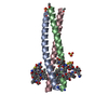
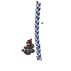
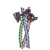
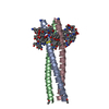
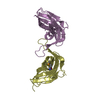
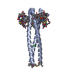
- PDB-2r3c: Structure of the gp41 N-peptide in complex with the HIV entry inh... -
+ Open data

- Basic information
| Entry | Database: PDB / ID: 2r3c | ||||||
|---|---|---|---|---|---|---|---|
| Title | Structure of the gp41 N-peptide in complex with the HIV entry inhibitor PIE1 | ||||||
 Components Components |
| ||||||
 Keywords Keywords |  VIRAL PROTEIN/VIRAL PROTEIN INHIBITOR / HIV / inhibitor / viral entry / PIE / VIRAL PROTEIN / VIRAL PROTEIN-VIRAL PROTEIN INHIBITOR complex VIRAL PROTEIN/VIRAL PROTEIN INHIBITOR / HIV / inhibitor / viral entry / PIE / VIRAL PROTEIN / VIRAL PROTEIN-VIRAL PROTEIN INHIBITOR complex | ||||||
| Function / homology | Single alpha-helices involved in coiled-coils or other helix-helix interfaces - #170 / Single alpha-helices involved in coiled-coils or other helix-helix interfaces / Up-down Bundle / Mainly Alpha / HIV entry inhibitor PIE1 / YTTRIUM (III) ION / polypeptide(D) / polypeptide(D) (> 10) Function and homology information Function and homology information | ||||||
| Biological species | synthetic construct (others) | ||||||
| Method | X-RAY DIFFRACTION / SYNCHROTRON / MOLECULAR REPLACEMENT / molecular replacement / Resolution: 1.73 Å | ||||||
 Authors Authors | VanDemark, A.P. / Welch, B. / Heroux, A. / Hill, C.P. / Kay, M.S. | ||||||
 Citation Citation | Journal: Proc.Natl.Acad.Sci.Usa / Year: 2007 Title: Potent D-peptide inhibitors of HIV-1 entry Authors: Welch, B.D. / Vandemark, A.P. / Heroux, A. / Hill, C.P. / Kay, M.S. | ||||||
| History |
|
- Structure visualization
| Structure viewer | Molecule: MolmilJmol/JSmol |
|---|
- Downloads & links
-Download
| PDBx/mmCIF format | 2r3c.cif.gz | 40.2 KB | Display | PDBx/mmCIF format |
|---|---|---|---|---|
| PDB format | pdb2r3c.ent.gz | 32.1 KB | Display | PDB format |
| PDBx/mmJSON format | 2r3c.json.gz | Tree view | PDBx/mmJSON format | |
| Others |  Other downloads Other downloads |
-Validation report
| Arichive directory | https://data.pdbj.org/pub/pdb/validation_reports/r3/2r3cftp://data.pdbj.org/pub/pdb/validation_reports/r3/2r3c | HTTPS FTP |
|---|
-Related structure data


-Links
 PDBj
PDBj


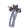
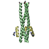
- Assembly
| Deposited unit | 
| |||||||||||||||
|---|---|---|---|---|---|---|---|---|---|---|---|---|---|---|---|---|
| 1 | 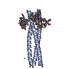
| |||||||||||||||
| 2 | 
| |||||||||||||||
| Unit cell |
| |||||||||||||||
| Components on special symmetry positions |
|
-Components
| #1: Protein/peptide | Mass: 5466.574 Da / Num. of mol.: 2 / Source method: obtained synthetically / Details: peptide synthesis / Source: (synth.) synthetic construct (others) #2: Polypeptide(D) |   Type: Peptide-like / Class: Inhibitor / Mass: 1831.103 Da / Num. of mol.: 2 / Source method: obtained synthetically / Details: peptide synthesis / Source: (synth.) synthetic construct (others) / References: HIV entry inhibitor PIE1 Type: Peptide-like / Class: Inhibitor / Mass: 1831.103 Da / Num. of mol.: 2 / Source method: obtained synthetically / Details: peptide synthesis / Source: (synth.) synthetic construct (others) / References: HIV entry inhibitor PIE1#3: Chemical | ChemComp-YT3 / Yttrium  Mass: 88.906 Da / Num. of mol.: 5 / Source method: obtained synthetically / Formula: Y Mass: 88.906 Da / Num. of mol.: 5 / Source method: obtained synthetically / Formula: Y#4: Chemical | ChemComp-CL / | Chloride  Mass: 35.453 Da / Num. of mol.: 1 / Source method: obtained synthetically / Formula: Cl Mass: 35.453 Da / Num. of mol.: 1 / Source method: obtained synthetically / Formula: Cl#5: Water | ChemComp-HOH / | Water Mass: 18.015 Da / Num. of mol.: 210 / Source method: isolated from a natural source / Formula: H2O Mass: 18.015 Da / Num. of mol.: 210 / Source method: isolated from a natural source / Formula: H2O |
|---|
-Experimental details
-Experiment
| Experiment | Method: X-RAY DIFFRACTION / Number of used crystals: 1 |
|---|
- Sample preparation
| Crystal | Density Matthews: 2.99 Å3/Da / Density % sol: 58.92 % |
|---|---|
| Crystal grow | Temperature: 298 K / Method: vapor diffusion / pH: 4.6 Details: 27% PEG 2000 MME, 0.1M Sodium Acetate, 0.4M YCl3, pH 4.6, VAPOR DIFFUSION, temperature 298K |
-Data collection
| Diffraction | Mean temperature: 100 K |
|---|---|
| Diffraction source | Source: SYNCHROTRON / Site: NSLS  / Beamline: X26C / Wavelength: 1.07274 Å / Beamline: X26C / Wavelength: 1.07274 Å |
| Detector | Type: ADSC QUANTUM 4 / Detector: CCD / Date: Apr 8, 2004 |
| Radiation | Monochromator: GRAPHITE / Protocol: SINGLE WAVELENGTH / Monochromatic (M) / Laue (L): M / Scattering type: x-ray |
| Radiation wavelength | Wavelength: 1.07274 Å / Relative weight: 1 |
| Reflection twin | Type: hemihedral / Operator: h,-h-k,-l / Fraction: 0.326 |
| Reflection | Resolution: 1.73→50 Å / Num. all: 35168 / Num. obs: 33506 / % possible obs: 95.3 % / Observed criterion σ(F): 0 / Observed criterion σ(I): 0 / Redundancy: 5.6 % / Rmerge(I) obs: 0.094 / Χ2: 1.327 / Net I/σ(I): 14.5 |
| Reflection shell | Resolution: 1.73→1.79 Å / Redundancy: 3.4 % / Rmerge(I) obs: 0.481 / Num. unique all: 2716 / Χ2: 0.864 / % possible all: 77.5 |
-Phasing
| Phasing | Method: molecular replacement |
|---|
- Processing
| Software |
| ||||||||||||||||||||||||||||
|---|---|---|---|---|---|---|---|---|---|---|---|---|---|---|---|---|---|---|---|---|---|---|---|---|---|---|---|---|---|
| Refinement | Method to determine structure: MOLECULAR REPLACEMENT / Resolution: 1.73→50 Å / Isotropic thermal model: isotropic / σ(F): 0 / σ(I): 0 / Stereochemistry target values: Engh & Huber / Details: Data has a twin fraction of 0.326
| ||||||||||||||||||||||||||||
| Solvent computation | Bsol: 53.063 Å2 | ||||||||||||||||||||||||||||
| Displacement parameters | Biso mean: 30.006 Å2
| ||||||||||||||||||||||||||||
| Refinement step | Cycle: LAST / Resolution: 1.73→50 Å
| ||||||||||||||||||||||||||||
| Refine LS restraints |
| ||||||||||||||||||||||||||||
| Xplor file |
|
