 Movie
Movie Controller

[English] 日本語
 Yorodumi
- PDB-4q4k: Crystal structure of nitronate monooxygenase from Pseudomonas aer... -
+ Open data

- Basic information
| Entry | Database: PDB / ID: 4q4k | ||||||
|---|---|---|---|---|---|---|---|
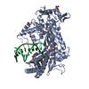
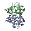
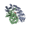
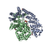
| Title | Crystal structure of nitronate monooxygenase from Pseudomonas aeruginosa PAO1 | ||||||
 Components Components | Nitronate Monooxygenase | ||||||
 Keywords Keywords | OXIDOREDUCTASE / TIM barrel / nitronate monooxygenase / FMN binding | ||||||
| Function / homology |  Function and homology informationOxidoreductases; Acting on single donors with incorporation of molecular oxygen (oxygenases); With incorporation of one atom of oxygen (internal monooxygenases or internal mixed-function oxidases) / nitronate monooxygenase activity / response to toxic substance / nucleotide binding Function and homology informationOxidoreductases; Acting on single donors with incorporation of molecular oxygen (oxygenases); With incorporation of one atom of oxygen (internal monooxygenases or internal mixed-function oxidases) / nitronate monooxygenase activity / response to toxic substance / nucleotide bindingSimilarity search - Function | ||||||
| Biological species |   Pseudomonas aeruginosa (bacteria) Pseudomonas aeruginosa (bacteria) | ||||||
| Method | X-RAY DIFFRACTION / SYNCHROTRON / MOLECULAR REPLACEMENT / Resolution: 1.44 Å | ||||||
 Authors Authors | Salvi, F. / Agniswamy, J. / Gadda, G. / Weber, I.T. | ||||||
 Citation Citation | Journal: J.Biol.Chem. / Year: 2014 Title: The Combined Structural and Kinetic Characterization of a Bacterial Nitronate Monooxygenase from Pseudomonas aeruginosa PAO1 Establishes NMO Class I and II. Authors: Salvi, F. / Agniswamy, J. / Yuan, H. / Vercammen, K. / Pelicaen, R. / Cornelis, P. / Spain, J.C. / Weber, I.T. / Gadda, G. | ||||||
| History |
|
- Structure visualization
| Structure viewer | Molecule: MolmilJmol/JSmol |
|---|
- Downloads & links
-Download
| PDBx/mmCIF format | 4q4k.cif.gz | 155.7 KB | Display | PDBx/mmCIF format |
|---|---|---|---|---|
| PDB format | pdb4q4k.ent.gz | 122.6 KB | Display | PDB format |
| PDBx/mmJSON format | 4q4k.json.gz | Tree view | PDBx/mmJSON format | |
| Others |  Other downloads Other downloads |
-Validation report
| Arichive directory | https://data.pdbj.org/pub/pdb/validation_reports/q4/4q4kftp://data.pdbj.org/pub/pdb/validation_reports/q4/4q4k | HTTPS FTP |
|---|
-Related structure data
| Similar structure data |
|---|


-Links
 PDBj
PDBj- Assembly
| Deposited unit | 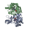
| ||||||||
|---|---|---|---|---|---|---|---|---|---|
| 1 |
| ||||||||
| Unit cell |
|
-Components
| #1: Protein | Mass: 37650.148 Da / Num. of mol.: 2 Source method: isolated from a genetically manipulated source Source: (gene. exp.) Pseudomonas aeruginosa (bacteria) / Strain: PAO1 / Gene: PA4202 / Plasmid: pET21a(+) / Production host: Escherichia coli (E. coli) / Strain (production host): Rosetta (DE3) pLysS / References: UniProt: Q9HWH9, nitronate monooxygenase#2: Chemical | Flavin mononucleotide  Mass: 456.344 Da / Num. of mol.: 2 / Source method: obtained synthetically / Formula: C17H21N4O9P Mass: 456.344 Da / Num. of mol.: 2 / Source method: obtained synthetically / Formula: C17H21N4O9P#3: Water | ChemComp-HOH / | Water Mass: 18.015 Da / Num. of mol.: 594 / Source method: isolated from a natural source / Formula: H2O Mass: 18.015 Da / Num. of mol.: 594 / Source method: isolated from a natural source / Formula: H2O |
|---|
-Experimental details
-Experiment
| Experiment | Method: X-RAY DIFFRACTION / Number of used crystals: 1 |
|---|
- Sample preparation
| Crystal | Density Matthews: 2.23 Å3/Da / Density % sol: 44.95 % |
|---|---|
| Crystal grow | Temperature: 298 K / pH: 7 Details: 14% PEG 5000 monomethylether, 0.1M HEPES-Na, pH 7.0, VAPOR DIFFUSION, HANGING DROP, temperature 298K |
-Data collection
| Diffraction | Mean temperature: 100 K |
|---|---|
| Diffraction source | Source: SYNCHROTRON / Site: APS  / Beamline: 22-BM / Wavelength: 0.8 / Beamline: 22-BM / Wavelength: 0.8 |
| Detector | Type: MARMOSAIC 225 mm CCD / Detector: CCD / Date: Oct 17, 2013 |
| Radiation | Monochromator: SI 220 / Protocol: SINGLE WAVELENGTH / Monochromatic (M) / Laue (L): M / Scattering type: x-ray |
| Radiation wavelength | Wavelength: 0.8 Å / Relative weight: 1 |
| Reflection | Resolution: 1.43→50 Å / Num. obs: 115873 / % possible obs: 94.1 % / Observed criterion σ(I): 2 |
| Reflection shell | Resolution: 1.43→1.48 Å / Redundancy: 3.5 % / Rmerge(I) obs: 0.456 / Mean I/σ(I) obs: 3.04 / % possible all: 68.9 |
- Processing
| Software |
| ||||||||||||||||||||||||||||||||||||||||||||||||||||||||||||||||||||||||||||||||||||||||||||||||||||||||||||||||||||||||||||||||||||||||||||||||||||||||||||||||||||||||||||||||||||||
|---|---|---|---|---|---|---|---|---|---|---|---|---|---|---|---|---|---|---|---|---|---|---|---|---|---|---|---|---|---|---|---|---|---|---|---|---|---|---|---|---|---|---|---|---|---|---|---|---|---|---|---|---|---|---|---|---|---|---|---|---|---|---|---|---|---|---|---|---|---|---|---|---|---|---|---|---|---|---|---|---|---|---|---|---|---|---|---|---|---|---|---|---|---|---|---|---|---|---|---|---|---|---|---|---|---|---|---|---|---|---|---|---|---|---|---|---|---|---|---|---|---|---|---|---|---|---|---|---|---|---|---|---|---|---|---|---|---|---|---|---|---|---|---|---|---|---|---|---|---|---|---|---|---|---|---|---|---|---|---|---|---|---|---|---|---|---|---|---|---|---|---|---|---|---|---|---|---|---|---|---|---|---|---|
| Refinement | Method to determine structure: MOLECULAR REPLACEMENT / Resolution: 1.44→37.6 Å / Cor.coef. Fo:Fc: 0.966 / Cor.coef. Fo:Fc free: 0.949 / Cross valid method: THROUGHOUT / ESU R: 0.08 / ESU R Free: 0.084 / Stereochemistry target values: MAXIMUM LIKELIHOOD / Details: HYDROGENS HAVE BEEN USED IF PRESENT IN THE INPUT
| ||||||||||||||||||||||||||||||||||||||||||||||||||||||||||||||||||||||||||||||||||||||||||||||||||||||||||||||||||||||||||||||||||||||||||||||||||||||||||||||||||||||||||||||||||||||
| Solvent computation | Ion probe radii: 0.8 Å / Shrinkage radii: 0.8 Å / VDW probe radii: 1.2 Å / Solvent model: MASK | ||||||||||||||||||||||||||||||||||||||||||||||||||||||||||||||||||||||||||||||||||||||||||||||||||||||||||||||||||||||||||||||||||||||||||||||||||||||||||||||||||||||||||||||||||||||
| Displacement parameters | Biso mean: 25.848 Å2
| ||||||||||||||||||||||||||||||||||||||||||||||||||||||||||||||||||||||||||||||||||||||||||||||||||||||||||||||||||||||||||||||||||||||||||||||||||||||||||||||||||||||||||||||||||||||
| Refinement step | Cycle: LAST / Resolution: 1.44→37.6 Å
| ||||||||||||||||||||||||||||||||||||||||||||||||||||||||||||||||||||||||||||||||||||||||||||||||||||||||||||||||||||||||||||||||||||||||||||||||||||||||||||||||||||||||||||||||||||||
| Refine LS restraints |
|