 Movie
Movie Controller

[English] 日本語
 Yorodumi
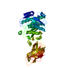
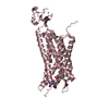
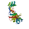
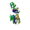
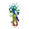
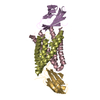
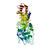
- PDB-4pyz: Crystal structure of the first two Ubl domains of Deubiquitylase USP7 -
+ Open data

- Basic information
| Entry | Database: PDB / ID: 4pyz | ||||||
|---|---|---|---|---|---|---|---|
| Title | Crystal structure of the first two Ubl domains of Deubiquitylase USP7 | ||||||
 Components Components | Ubiquitin carboxyl-terminal hydrolase 7 | ||||||
 Keywords Keywords |  HYDROLASE / Deubiquitylase / ubiquitin-like domain / Structural Genomics / Structural Genomics Consortium / SGC HYDROLASE / Deubiquitylase / ubiquitin-like domain / Structural Genomics / Structural Genomics Consortium / SGC | ||||||
| Function / homology |  Function and homology information Function and homology informationregulation of telomere capping / monoubiquitinated protein deubiquitination / regulation of retrograde transport, endosome to Golgi / deubiquitinase activity / DNA alkylation repair / regulation of DNA-binding transcription factor activity / negative regulation of gene expression via chromosomal CpG island methylation / K48-linked deubiquitinase activity / symbiont-mediated disruption of host cell PML body / negative regulation of NF-kappaB transcription factor activity ...regulation of telomere capping / monoubiquitinated protein deubiquitination / regulation of retrograde transport, endosome to Golgi / deubiquitinase activity / DNA alkylation repair / regulation of DNA-binding transcription factor activity / negative regulation of gene expression via chromosomal CpG island methylation / K48-linked deubiquitinase activity / symbiont-mediated disruption of host cell PML body / negative regulation of NF-kappaB transcription factor activity / protein deubiquitination / negative regulation of proteasomal ubiquitin-dependent protein catabolic process / transcription-coupled nucleotide-excision repair / negative regulation of gluconeogenesis / negative regulation of TORC1 signaling / Regulation of PTEN localization / Synthesis of active ubiquitin: roles of E1 and E2 enzymes / regulation of signal transduction by p53 class mediator / regulation of protein stability / regulation of circadian rhythm / Transcription-Coupled Nucleotide Excision Repair (TC-NER) / Formation of TC-NER Pre-Incision Complex / PML body / Dual incision in TC-NER / Gap-filling DNA repair synthesis and ligation in TC-NER / Regulation of TP53 Degradation / rhythmic process / p53 binding / chromosome / ubiquitinyl hydrolase 1 / cysteine-type deubiquitinase activity / nuclear body / protein stabilization / protein ubiquitination / Ub-specific processing proteases / cysteine-type endopeptidase activity / protein-containing complex / proteolysis / nucleoplasm / nucleus / cytosolSimilarity search - Function | ||||||
| Biological species |  Homo sapiens (human) Homo sapiens (human) | ||||||
| Method | X-RAY DIFFRACTION / SYNCHROTRON / MOLECULAR REPLACEMENT / Resolution: 2.84 Å | ||||||
 Authors Authors | Walker, J.R. / Dong, A. / Ong, M.S. / Dhe-Paganon, S. / Kania, J. / Bountra, C. / Arrowsmith, C.H. / Edwards, A.M. / Tong, Y. / Structural Genomics Consortium (SGC) | ||||||
 Citation Citation | Journal: to be published Title: Crystal structure of the first two Ubl domains of Deubiquitylase USP7 Authors: Ong, M.S. / Walker, J.R. / Dong, A. / Dhe-Paganon, S. / Kania, J. / Bountra, C. / Arrowsmith, C.H. / Edwards, A.M. / Tong, Y. / Structural Genomics Consortium (SGC) | ||||||
| History |
|
- Structure visualization
| Structure viewer | Molecule: MolmilJmol/JSmol |
|---|
- Downloads & links
-Download
| PDBx/mmCIF format | 4pyz.cif.gz | 216.7 KB | Display | PDBx/mmCIF format |
|---|---|---|---|---|
| PDB format | pdb4pyz.ent.gz | 174.6 KB | Display | PDB format |
| PDBx/mmJSON format | 4pyz.json.gz | Tree view | PDBx/mmJSON format | |
| Others |  Other downloads Other downloads |
-Validation report
| Arichive directory | https://data.pdbj.org/pub/pdb/validation_reports/py/4pyzftp://data.pdbj.org/pub/pdb/validation_reports/py/4pyz | HTTPS FTP |
|---|
-Related structure data
| Related structure data | 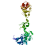 2ylmS S: Starting model for refinement |
|---|---|
| Similar structure data |

-Links
 PDBj
PDBj


- Assembly
| Deposited unit | 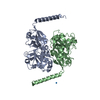
| ||||||||
|---|---|---|---|---|---|---|---|---|---|
| 1 | 
| ||||||||
| 2 | 
| ||||||||
| Unit cell |
| ||||||||
| Details | AS PER THE AUTHORS THE BIOLOGICAL ASSEMBLY IS UNKNOWN |
-Components
| #1: Protein | Mass: 31733.713 Da / Num. of mol.: 2 / Fragment: UNP residues 537-793 Source method: isolated from a genetically manipulated source Source: (gene. exp.) Homo sapiens (human) / Gene: USP7, HAUSP / Plasmid: pET28-LIC / Production host:  Escherichia coli (E. coli) / Strain (production host): BL21(DE3)-V2R / References: UniProt: Q93009, ubiquitinyl hydrolase 1 Escherichia coli (E. coli) / Strain (production host): BL21(DE3)-V2R / References: UniProt: Q93009, ubiquitinyl hydrolase 1#2: Chemical |   Num. of mol.: 2 / Source method: obtained synthetically Num. of mol.: 2 / Source method: obtained synthetically#3: Water | ChemComp-HOH / | Water Mass: 18.015 Da / Num. of mol.: 12 / Source method: isolated from a natural source / Formula: H2O Mass: 18.015 Da / Num. of mol.: 12 / Source method: isolated from a natural source / Formula: H2O |
|---|
-Experimental details
-Experiment
| Experiment | Method: X-RAY DIFFRACTION / Number of used crystals: 1 |
|---|
- Sample preparation
| Crystal | Density Matthews: 3.24 Å3/Da / Density % sol: 62.09 % |
|---|---|
| Crystal grow | Temperature: 291 K / Method: vapor diffusion, hanging drop / pH: 5.6 Details: 15% PEG 4000, 0.2 M NH4Ac, 0.1 M NaCitrate pH5.6, vapor diffusion hanging drop, temperature 291K |
-Data collection
| Diffraction | Mean temperature: 100 K | |||||||||||||||||||||||||||||||||||||||||||||||||||||||||||||||||||||||||||||||||||||||||||||||||||||||||||||||||||||||||||||||||||||||||||||||||||
|---|---|---|---|---|---|---|---|---|---|---|---|---|---|---|---|---|---|---|---|---|---|---|---|---|---|---|---|---|---|---|---|---|---|---|---|---|---|---|---|---|---|---|---|---|---|---|---|---|---|---|---|---|---|---|---|---|---|---|---|---|---|---|---|---|---|---|---|---|---|---|---|---|---|---|---|---|---|---|---|---|---|---|---|---|---|---|---|---|---|---|---|---|---|---|---|---|---|---|---|---|---|---|---|---|---|---|---|---|---|---|---|---|---|---|---|---|---|---|---|---|---|---|---|---|---|---|---|---|---|---|---|---|---|---|---|---|---|---|---|---|---|---|---|---|---|---|---|---|
| Diffraction source | Source: SYNCHROTRON / Site: APS  / Beamline: 19-ID / Wavelength: 0.97924 Å / Beamline: 19-ID / Wavelength: 0.97924 Å | |||||||||||||||||||||||||||||||||||||||||||||||||||||||||||||||||||||||||||||||||||||||||||||||||||||||||||||||||||||||||||||||||||||||||||||||||||
| Detector | Type: ADSC QUANTUM 315r / Detector: CCD / Date: Oct 20, 2013 | |||||||||||||||||||||||||||||||||||||||||||||||||||||||||||||||||||||||||||||||||||||||||||||||||||||||||||||||||||||||||||||||||||||||||||||||||||
| Radiation | Protocol: SINGLE WAVELENGTH / Monochromatic (M) / Laue (L): M / Scattering type: x-ray | |||||||||||||||||||||||||||||||||||||||||||||||||||||||||||||||||||||||||||||||||||||||||||||||||||||||||||||||||||||||||||||||||||||||||||||||||||
| Radiation wavelength | Wavelength: 0.97924 Å / Relative weight: 1 | |||||||||||||||||||||||||||||||||||||||||||||||||||||||||||||||||||||||||||||||||||||||||||||||||||||||||||||||||||||||||||||||||||||||||||||||||||
| Reflection | Resolution: 2.83→50 Å / Num. obs: 19822 / % possible obs: 99.3 % / Redundancy: 6.8 % / Biso Wilson estimate: 82.16 Å2 / Rmerge(I) obs: 0.095 / Χ2: 1.109 / Net I/σ(I): 22.2 | |||||||||||||||||||||||||||||||||||||||||||||||||||||||||||||||||||||||||||||||||||||||||||||||||||||||||||||||||||||||||||||||||||||||||||||||||||
| Reflection shell |
|
- Processing
| Software |
| ||||||||||||||||||||||||||||||||||||||||||||||||||||||||||||||||||||||||||||||||||||||||||||||||||||||||||||
|---|---|---|---|---|---|---|---|---|---|---|---|---|---|---|---|---|---|---|---|---|---|---|---|---|---|---|---|---|---|---|---|---|---|---|---|---|---|---|---|---|---|---|---|---|---|---|---|---|---|---|---|---|---|---|---|---|---|---|---|---|---|---|---|---|---|---|---|---|---|---|---|---|---|---|---|---|---|---|---|---|---|---|---|---|---|---|---|---|---|---|---|---|---|---|---|---|---|---|---|---|---|---|---|---|---|---|---|---|---|
| Refinement | Method to determine structure: MOLECULAR REPLACEMENT Starting model: PDB entry 2YLM Resolution: 2.84→40.71 Å / Cor.coef. Fo:Fc: 0.8741 / Cor.coef. Fo:Fc free: 0.8748 / SU R Cruickshank DPI: 0.768 / Cross valid method: THROUGHOUT / σ(F): 0 / Stereochemistry target values: Engh & Huber
| ||||||||||||||||||||||||||||||||||||||||||||||||||||||||||||||||||||||||||||||||||||||||||||||||||||||||||||
| Displacement parameters | Biso max: 147.3 Å2 / Biso mean: 80.95 Å2 / Biso min: 30 Å2
| ||||||||||||||||||||||||||||||||||||||||||||||||||||||||||||||||||||||||||||||||||||||||||||||||||||||||||||
| Refine analyze | Luzzati coordinate error obs: 0.564 Å | ||||||||||||||||||||||||||||||||||||||||||||||||||||||||||||||||||||||||||||||||||||||||||||||||||||||||||||
| Refinement step | Cycle: LAST / Resolution: 2.84→40.71 Å
| ||||||||||||||||||||||||||||||||||||||||||||||||||||||||||||||||||||||||||||||||||||||||||||||||||||||||||||
| Refine LS restraints |
| ||||||||||||||||||||||||||||||||||||||||||||||||||||||||||||||||||||||||||||||||||||||||||||||||||||||||||||
| LS refinement shell | Resolution: 2.84→2.99 Å / Total num. of bins used: 10
| ||||||||||||||||||||||||||||||||||||||||||||||||||||||||||||||||||||||||||||||||||||||||||||||||||||||||||||
| Refinement TLS params. | Method: refined / Refine-ID: X-RAY DIFFRACTION
| ||||||||||||||||||||||||||||||||||||||||||||||||||||||||||||||||||||||||||||||||||||||||||||||||||||||||||||
| Refinement TLS group |
|
