 Movie
Movie Controller


+ Open data

- Basic information
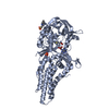
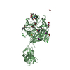
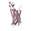
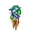
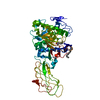
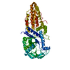
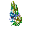
| Entry | Database: PDB / ID: 6syn | ||||||
|---|---|---|---|---|---|---|---|
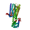
| Title | Crystal structure of Y. pestis penicillin-binding protein 3 | ||||||
 Components Components | Peptidoglycan D,D-transpeptidase FtsI | ||||||
 Keywords Keywords |  CYTOSOLIC PROTEIN / Class B PBP / Yersinia pestis / HMM transpeptidase / periplasmic protein CYTOSOLIC PROTEIN / Class B PBP / Yersinia pestis / HMM transpeptidase / periplasmic protein | ||||||
| Function / homology |  Function and homology informationpeptidoglycan glycosyltransferase activity / serine-type D-Ala-D-Ala carboxypeptidase / FtsZ-dependent cytokinesis / serine-type D-Ala-D-Ala carboxypeptidase activity / division septum assembly / plasma membrane => GO:0005886 / penicillin binding / peptidoglycan biosynthetic process / cell wall organization / regulation of cell shape ...peptidoglycan glycosyltransferase activity / serine-type D-Ala-D-Ala carboxypeptidase / FtsZ-dependent cytokinesis / serine-type D-Ala-D-Ala carboxypeptidase activity / division septum assembly / plasma membrane => GO:0005886 / penicillin binding / peptidoglycan biosynthetic process / cell wall organization / regulation of cell shape / membrane => GO:0016020 / proteolysis / plasma membrane Function and homology informationpeptidoglycan glycosyltransferase activity / serine-type D-Ala-D-Ala carboxypeptidase / FtsZ-dependent cytokinesis / serine-type D-Ala-D-Ala carboxypeptidase activity / division septum assembly / plasma membrane => GO:0005886 / penicillin binding / peptidoglycan biosynthetic process / cell wall organization / regulation of cell shape ...peptidoglycan glycosyltransferase activity / serine-type D-Ala-D-Ala carboxypeptidase / FtsZ-dependent cytokinesis / serine-type D-Ala-D-Ala carboxypeptidase activity / division septum assembly / plasma membrane => GO:0005886 / penicillin binding / peptidoglycan biosynthetic process / cell wall organization / regulation of cell shape / membrane => GO:0016020 / proteolysis / plasma membraneSimilarity search - Function | ||||||
| Biological species |   Yersinia pestis (bacteria) Yersinia pestis (bacteria) | ||||||
| Method | X-RAY DIFFRACTION / MOLECULAR REPLACEMENT / Resolution: 2.63 Å | ||||||
 Authors Authors | Pankov, G. / Hunter, W.N. / Dawson, A. | ||||||
 Citation Citation | Journal: To Be Published Title: The structure of penicillin-binding protein 3 from Yersinia pestis Authors: Pankov, G. | ||||||
| History |
|
- Structure visualization
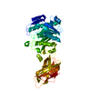
| Structure viewer | Molecule: MolmilJmol/JSmol |
|---|
- Downloads & links
-Download
| PDBx/mmCIF format | 6syn.cif.gz | 108 KB | Display | PDBx/mmCIF format |
|---|---|---|---|---|
| PDB format | pdb6syn.ent.gz | 79.2 KB | Display | PDB format |
| PDBx/mmJSON format | 6syn.json.gz | Tree view | PDBx/mmJSON format | |
| Others |  Other downloads Other downloads |
-Validation report
| Arichive directory | https://data.pdbj.org/pub/pdb/validation_reports/sy/6synftp://data.pdbj.org/pub/pdb/validation_reports/sy/6syn | HTTPS FTP |
|---|
-Related structure data
| Related structure data | 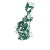 6tudC 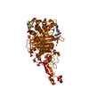 4bjpS S: Starting model for refinement C: citing same article ( |
|---|---|
| Similar structure data |
-Links
 PDBj
PDBj
- Assembly
| Deposited unit | 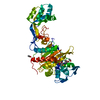
| ||||||||
|---|---|---|---|---|---|---|---|---|---|
| 1 |
| ||||||||
| Unit cell |
|
-Components
| #1: Protein | Mass: 57646.836 Da / Num. of mol.: 1 Source method: isolated from a genetically manipulated source Source: (gene. exp.) Yersinia pestis (bacteria) / Gene: ftsI, YPO0549 / Production host: Escherichia coli (E. coli)References: UniProt: Q0WJB8, UniProt: A0A3N4B5A3*PLUS, serine-type D-Ala-D-Ala carboxypeptidase | ||||||
|---|---|---|---|---|---|---|---|
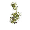
| #2: Chemical | Acetate  Mass: 59.044 Da / Num. of mol.: 2 / Source method: obtained synthetically / Formula: C2H3O2 Mass: 59.044 Da / Num. of mol.: 2 / Source method: obtained synthetically / Formula: C2H3O2#3: Chemical | ChemComp-CB9 / ( |   Mass: 380.416 Da / Num. of mol.: 1 / Source method: obtained synthetically / Formula: C17H20N2O6S Mass: 380.416 Da / Num. of mol.: 1 / Source method: obtained synthetically / Formula: C17H20N2O6S#4: Water | ChemComp-HOH / | Water Mass: 18.015 Da / Num. of mol.: 144 / Source method: isolated from a natural source / Formula: H2O Mass: 18.015 Da / Num. of mol.: 144 / Source method: isolated from a natural source / Formula: H2OHas ligand of interest | Y | |
-Experimental details
-Experiment
| Experiment | Method: X-RAY DIFFRACTION / Number of used crystals: 1 |
|---|
- Sample preparation
| Crystal | Density Matthews: 2.04 Å3/Da / Density % sol: 40 % / Description: rectangular prisms |
|---|---|
| Crystal grow | Temperature: 296 K / Method: vapor diffusion, sitting drop Details: 0.2 uL of protein (in 20 mM Tris-HCl pH 7.5 150 mM NaCl and 2 mM carbenicillin) at 6.7 mg/ml and 0.2 uL of precipitant (0.2 M magnesium acetate, 0.1 M sodium cacodylate pH 6.5 and 20% PEG 8000) |
-Data collection
| Diffraction | Mean temperature: 100 K / Serial crystal experiment: N |
|---|---|
| Diffraction source | Source: ROTATING ANODE / Type: RIGAKU MICROMAX-007 HF / Wavelength: 1.54178 Å |
| Detector | Type: RIGAKU SATURN 944+ / Detector: CCD / Date: Jan 16, 2017 |
| Radiation | Protocol: SINGLE WAVELENGTH / Monochromatic (M) / Laue (L): M / Scattering type: x-ray |
| Radiation wavelength | Wavelength: 1.54178 Å / Relative weight: 1 |
| Reflection | Resolution: 2.63→36 Å / Num. obs: 14613 / % possible obs: 98.7 % / Redundancy: 3.9 % / Biso Wilson estimate: 8.1 Å2 / CC1/2: 0.971 / Rmerge(I) obs: 0.122 / Rpim(I) all: 0.097 / Rrim(I) all: 0.153 / Net I/σ(I): 7.8 |
| Reflection shell | Resolution: 2.63→2.74 Å / Redundancy: 3.1 % / Rmerge(I) obs: 0.234 / Mean I/σ(I) obs: 3.9 / Num. unique obs: 1663 / CC1/2: 0.652 / Rpim(I) all: 0.203 / Rrim(I) all: 0.312 / % possible all: 93.8 |
- Processing
| Software |
| ||||||||||||||||||||||||||||||||||||||||||||||||||||||||||||
|---|---|---|---|---|---|---|---|---|---|---|---|---|---|---|---|---|---|---|---|---|---|---|---|---|---|---|---|---|---|---|---|---|---|---|---|---|---|---|---|---|---|---|---|---|---|---|---|---|---|---|---|---|---|---|---|---|---|---|---|---|---|
| Refinement | Method to determine structure: MOLECULAR REPLACEMENT Starting model: 4BJP Resolution: 2.63→36 Å / Cor.coef. Fo:Fc: 0.895 / Cor.coef. Fo:Fc free: 0.819 / SU B: 13.841 / SU ML: 0.288 / Cross valid method: THROUGHOUT / σ(F): 0 / ESU R Free: 0.382 Details: HYDROGENS HAVE BEEN ADDED IN THE RIDING POSITIONS U VALUES : REFINED INDIVIDUALLY
| ||||||||||||||||||||||||||||||||||||||||||||||||||||||||||||
| Solvent computation | Ion probe radii: 0.8 Å / Shrinkage radii: 0.8 Å / VDW probe radii: 1.2 Å | ||||||||||||||||||||||||||||||||||||||||||||||||||||||||||||
| Displacement parameters | Biso max: 62 Å2 / Biso mean: 15.899 Å2 / Biso min: 5 Å2
| ||||||||||||||||||||||||||||||||||||||||||||||||||||||||||||
| Refinement step | Cycle: final / Resolution: 2.63→36 Å
| ||||||||||||||||||||||||||||||||||||||||||||||||||||||||||||
| Refine LS restraints |
| ||||||||||||||||||||||||||||||||||||||||||||||||||||||||||||
| LS refinement shell | Resolution: 2.63→2.696 Å / Rfactor Rfree error: 0
|
