 Movie
Movie Controller


+ Open data

- Basic information
| Entry | Database: PDB / ID: 4pyk | ||||||
|---|---|---|---|---|---|---|---|
| Title | human COMT, double domain swap | ||||||
 Components Components | Catechol O-methyltransferase Catechol-O-methyltransferase Catechol-O-methyltransferase | ||||||
 Keywords Keywords | TRANSFERASE / METHYLTRANSFERASE / NEUROTRANSMITTER DEGRADATION / CONFORMATIONAL CHANGE / CATECHOLAMINE METABOLISM / CELL MEMBRANE / MAGNESIUM / MEMBRANE / METAL-BINDING / S-ADENOSYL-L-METHIONINE / SIGNAL-ANCHOR / ENZYME | ||||||
| Function / homology |  Function and homology information Function and homology informationEnzymatic degradation of dopamine by COMT / Enzymatic degradation of Dopamine by monoamine oxidase / norepinephrine secretion / response to dopamine / mastication / catecholamine catabolic process / catechol O-methyltransferase activity / renal sodium excretion / : / : ...Enzymatic degradation of dopamine by COMT / Enzymatic degradation of Dopamine by monoamine oxidase / norepinephrine secretion / response to dopamine / mastication / catecholamine catabolic process / catechol O-methyltransferase activity / renal sodium excretion / : / : / catechol O-methyltransferase / developmental process / renal filtration / renin secretion into blood stream / dopamine secretion / Methylation / renal albumin absorption / response to salt / habituation / artery development / cerebellar cortex morphogenesis / cellular response to phosphate starvation / dopamine catabolic process / norepinephrine metabolic process / glomerulus development / synaptic transmission, dopaminergic / O-methyltransferase activity / response to angiotensin / cellular response to cocaine / exploration behavior / response to food / cholesterol efflux / response to corticosterone / dopamine metabolic process / glycogen metabolic process / prostaglandin metabolic process / startle response / detection of temperature stimulus involved in sensory perception of pain / : / behavioral fear response / response to amphetamine / methyltransferase activity / response to cytokine / visual learning / multicellular organism growth / response to toxic substance / memory / response to wounding / gene expression / methylation / response to oxidative stress / Potential therapeutics for SARS / response to hypoxia / response to xenobiotic stimulus / axon / intracellular membrane-bounded organelle / synapse / dendrite / magnesium ion binding / extracellular exosome / membrane / plasma membrane / cytosolSimilarity search - Function | ||||||
| Biological species |  Homo sapiens (human) Homo sapiens (human) | ||||||
| Method | X-RAY DIFFRACTION / SYNCHROTRON / MOLECULAR REPLACEMENT / Resolution: 2.22 Å | ||||||
 Authors Authors | Ehler, A. / Benz, J. / Schlatter, D. / Rudolph, M.G. | ||||||
 Citation Citation | Journal: Acta Crystallogr.,Sect.D / Year: 2014 Title: Mapping the conformational space accessible to catechol-O-methyltransferase. Authors: Ehler, A. / Benz, J. / Schlatter, D. / Rudolph, M.G. | ||||||
| History |
|
- Structure visualization
| Structure viewer | Molecule: MolmilJmol/JSmol |
|---|
- Downloads & links
-Download
| PDBx/mmCIF format | 4pyk.cif.gz | 99.5 KB | Display | PDBx/mmCIF format |
|---|---|---|---|---|
| PDB format | pdb4pyk.ent.gz | 75.9 KB | Display | PDB format |
| PDBx/mmJSON format | 4pyk.json.gz | Tree view | PDBx/mmJSON format | |
| Others |  Other downloads Other downloads |
-Validation report
| Arichive directory | https://data.pdbj.org/pub/pdb/validation_reports/py/4pykftp://data.pdbj.org/pub/pdb/validation_reports/py/4pyk | HTTPS FTP |
|---|
-Related structure data
| Related structure data |  4p7fC 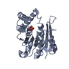 4p7gC  4p7jC 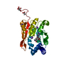 4p7kC  4pyiC 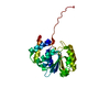 4pyjC 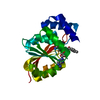 4pylC  4pymC  4pynC 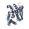 4pyoC 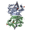 4pyqC C: citing same article ( |
|---|---|
| Similar structure data |


-Links
 PDBj
PDBj- Assembly
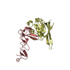
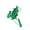
| Deposited unit | 
| ||||||||
|---|---|---|---|---|---|---|---|---|---|
| 1 |
| ||||||||
| 2 | 
| ||||||||
| Unit cell |
| ||||||||
| Components on special symmetry positions |
|
-Components
| #1: Protein | Catechol-O-methyltransferase Mass: 24478.076 Da / Num. of mol.: 1 / Fragment: unp residues 51-271 Source method: isolated from a genetically manipulated source Source: (gene. exp.) Homo sapiens (human) / Gene: COMT / Production host:  Escherichia coli (E. coli) / References: UniProt: P21964, catechol O-methyltransferase Escherichia coli (E. coli) / References: UniProt: P21964, catechol O-methyltransferase |
|---|---|
| #2: Chemical | ChemComp-NA /   Mass: 22.990 Da / Num. of mol.: 1 / Source method: obtained synthetically / Formula: Na Mass: 22.990 Da / Num. of mol.: 1 / Source method: obtained synthetically / Formula: Na |
| #3: Chemical | ChemComp-MG /   Mass: 24.305 Da / Num. of mol.: 1 / Source method: obtained synthetically / Formula: Mg Mass: 24.305 Da / Num. of mol.: 1 / Source method: obtained synthetically / Formula: Mg |
| #4: Chemical | ChemComp-CL / Chloride  Mass: 35.453 Da / Num. of mol.: 1 / Source method: obtained synthetically / Formula: Cl Mass: 35.453 Da / Num. of mol.: 1 / Source method: obtained synthetically / Formula: Cl |
| #5: Water | ChemComp-HOH / Water Mass: 18.015 Da / Num. of mol.: 50 / Source method: isolated from a natural source / Formula: H2O Mass: 18.015 Da / Num. of mol.: 50 / Source method: isolated from a natural source / Formula: H2O |
-Experimental details
-Experiment
| Experiment | Method: X-RAY DIFFRACTION / Number of used crystals: 1 |
|---|
- Sample preparation
| Crystal | Density Matthews: 2.39 Å3/Da / Density % sol: 48.61 % |
|---|---|
| Crystal grow | Temperature: 293 K / pH: 7.5 Details: pH 7.5, VAPOR DIFFUSION, SITTING DROP, temperature 293K |
-Data collection
| Diffraction | Mean temperature: 100 K |
|---|---|
| Diffraction source | Source: SYNCHROTRON / Site: SLS  / Beamline: X10SA / Wavelength: 1 / Beamline: X10SA / Wavelength: 1 |
| Detector | Type: DECTRIS PILATUS 6M / Detector: PIXEL / Date: Feb 17, 2010 |
| Radiation | Protocol: SINGLE WAVELENGTH / Monochromatic (M) / Laue (L): M / Scattering type: x-ray |
| Radiation wavelength | Wavelength: 1 Å / Relative weight: 1 |
| Reflection | Resolution: 2.22→40.222 Å / Num. obs: 78926 / % possible obs: 99.8 % / Observed criterion σ(F): 0 / Observed criterion σ(I): 0 / Redundancy: 6.6 % / Rmerge(I) obs: 0.1041 / Net I/σ(I): 11.5483 |
| Reflection shell | Resolution: 2.22→2.3 Å / Redundancy: 6.6 % / Rmerge(I) obs: 1.17196 / Mean I/σ(I) obs: 1.5281 / % possible all: 99.8 |
- Processing
| Software |
| ||||||||||||||||||||||||||||||||||||||||||||||||||||||||||||||||||||||||||||||||||||||||||||||||||||
|---|---|---|---|---|---|---|---|---|---|---|---|---|---|---|---|---|---|---|---|---|---|---|---|---|---|---|---|---|---|---|---|---|---|---|---|---|---|---|---|---|---|---|---|---|---|---|---|---|---|---|---|---|---|---|---|---|---|---|---|---|---|---|---|---|---|---|---|---|---|---|---|---|---|---|---|---|---|---|---|---|---|---|---|---|---|---|---|---|---|---|---|---|---|---|---|---|---|---|---|---|---|
| Refinement | Method to determine structure: MOLECULAR REPLACEMENT Starting model: in house model Resolution: 2.22→40.222 Å / SU ML: 0.29 / σ(F): 22.8 / Phase error: 30.06 / Stereochemistry target values: ML
| ||||||||||||||||||||||||||||||||||||||||||||||||||||||||||||||||||||||||||||||||||||||||||||||||||||
| Solvent computation | Shrinkage radii: 0.9 Å / VDW probe radii: 1.11 Å / Solvent model: FLAT BULK SOLVENT MODEL | ||||||||||||||||||||||||||||||||||||||||||||||||||||||||||||||||||||||||||||||||||||||||||||||||||||
| Refinement step | Cycle: LAST / Resolution: 2.22→40.222 Å
| ||||||||||||||||||||||||||||||||||||||||||||||||||||||||||||||||||||||||||||||||||||||||||||||||||||
| Refine LS restraints |
| ||||||||||||||||||||||||||||||||||||||||||||||||||||||||||||||||||||||||||||||||||||||||||||||||||||
| LS refinement shell |
| ||||||||||||||||||||||||||||||||||||||||||||||||||||||||||||||||||||||||||||||||||||||||||||||||||||
| Refinement TLS params. | Method: refined / Refine-ID: X-RAY DIFFRACTION
| ||||||||||||||||||||||||||||||||||||||||||||||||||||||||||||||||||||||||||||||||||||||||||||||||||||
| Refinement TLS group |
|
