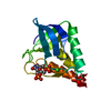
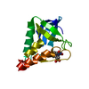
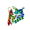
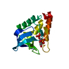
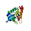
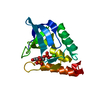
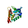
Crystal structure of Staphylcoccal nuclease variant Delta+PHS I92S at cryogenic temperature
Components
ThermonucleaseMicrococcal nuclease
Keywords
HYDROLASE / nuclease / hyperstable / pdTp / polar group
Function / homology
Function and homology information
endonuclease activity, active with either ribo- or deoxyribonucleic acids and producing 3'-phosphomonoesters / micrococcal nuclease / nucleic acid binding / extracellular region / membrane / metal ion binding Similarity search - Function
Thermonuclease family signature 1. / OB fold (Dihydrolipoamide Acetyltransferase, E2P) - #90 / Thermonuclease active site / Thermonuclease family signature 2. / Staphylococcal nuclease (SNase-like), OB-fold / Staphylococcal nuclease homologue / Thermonuclease domain profile. / Staphylococcal nuclease homologues / SNase-like, OB-fold superfamily / OB fold (Dihydrolipoamide Acetyltransferase, E2P) ...Thermonuclease family signature 1. / OB fold (Dihydrolipoamide Acetyltransferase, E2P) - #90 / Thermonuclease active site / Thermonuclease family signature 2. / Staphylococcal nuclease (SNase-like), OB-fold / Staphylococcal nuclease homologue / Thermonuclease domain profile. / Staphylococcal nuclease homologues / SNase-like, OB-fold superfamily / OB fold (Dihydrolipoamide Acetyltransferase, E2P) / Beta Barrel / Mainly Beta Similarity search - Domain/homology
Resolution: 1.8→37.94 Å / Cor.coef. Fo:Fc: 0.954 / Cor.coef. Fo:Fc free: 0.938 / WRfactor Rfree: 0.2116 / WRfactor Rwork: 0.1872 / FOM work R set: 0.8198 / SU B: 5.237 / SU ML: 0.089 / SU R Cruickshank DPI: 0.1392 / SU Rfree: 0.1272 / Data cutoff high absF: 0 / Data cutoff low absF: 0 / Cross valid method: THROUGHOUT / σ(F): 0 / ESU R: 0.139 / ESU R Free: 0.127 / Stereochemistry target values: MAXIMUM LIKELIHOOD / Details: HYDROGENS HAVE BEEN ADDED IN THE RIDING POSITIONS
Rfactor
Num. reflection
% reflection
Selection details
Rfree
0.2269
630
4.9 %
RANDOM
Rwork
0.1964
12320
-
-
obs
0.1979
12950
99.81 %
-
Solvent computation
Ion probe radii: 0.8 Å / Shrinkage radii: 0.8 Å / VDW probe radii: 1.2 Å / Solvent model: MASK
In the structure databanks used in Yorodumi, some data are registered as the other names, "COVID-19 virus" and "2019-nCoV". Here are the details of the virus and the list of structure data.
Jan 31, 2019. EMDB accession codes are about to change! (news from PDBe EMDB page)
EMDB accession codes are about to change! (news from PDBe EMDB page)
The allocation of 4 digits for EMDB accession codes will soon come to an end. Whilst these codes will remain in use, new EMDB accession codes will include an additional digit and will expand incrementally as the available range of codes is exhausted. The current 4-digit format prefixed with “EMD-” (i.e. EMD-XXXX) will advance to a 5-digit format (i.e. EMD-XXXXX), and so on. It is currently estimated that the 4-digit codes will be depleted around Spring 2019, at which point the 5-digit format will come into force.
The EM Navigator/Yorodumi systems omit the EMD- prefix.
Related info.:Q: What is EMD? / ID/Accession-code notation in Yorodumi/EM Navigator
Yorodumi is a browser for structure data from EMDB, PDB, SASBDB, etc.
This page is also the successor to EM Navigator detail page, and also detail information page/front-end page for Omokage search.
The word "yorodu" (or yorozu) is an old Japanese word meaning "ten thousand". "mi" (miru) is to see.
Related info.:EMDB / PDB / SASBDB / Comparison of 3 databanks / Yorodumi Search / Aug 31, 2016. New EM Navigator & Yorodumi / Yorodumi Papers / Jmol/JSmol / Function and homology information / Changes in new EM Navigator and Yorodumi
 Movie
Movie Controller

 Yorodumi
 Open data
 Basic information
 Components
 Micrococcal nuclease
  Keywords
 Function and homology information

 Authors
 United States, 1items
  Citation
 Structure visualization
 Downloads & links
 Other downloads
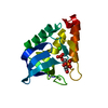


 PDBj
PDBj Assembly
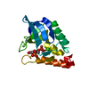

 Type: DNA linking / Mass: 402.188 Da / Num. of mol.: 1 / Source method: obtained synthetically / Formula: C10H16N2O11P2
 Mass: 40.078 Da / Num. of mol.: 1 / Source method: obtained synthetically / Formula: Ca
 Mass: 18.015 Da / Num. of mol.: 67 / Source method: isolated from a natural source / Formula: H2O
 Sample preparation
 Processing