 Movie
Movie Controller

[English] 日本語
 Yorodumi
- PDB-4kjn: Crystal structure of Staphylococcal nuclease variant Delta+PHS V2... -
+ Open data

- Basic information
| Entry | Database: PDB / ID: 4kjn | ||||||
|---|---|---|---|---|---|---|---|
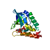
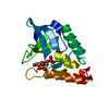
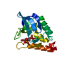
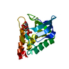
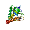
| Title | Crystal structure of Staphylococcal nuclease variant Delta+PHS V23T/V66A/V99T at cryogenic temperature | ||||||
 Components Components | Thermonuclease Micrococcal nuclease Micrococcal nuclease | ||||||
 Keywords Keywords | HYDROLASE / Staphylococcal nuclease / hyperstable variant / pdtp / cavity / pressure | ||||||
| Function / homology |  Function and homology information Function and homology informationendonuclease activity, active with either ribo- or deoxyribonucleic acids and producing 3'-phosphomonoesters / micrococcal nuclease / nucleic acid binding / extracellular region / membrane / metal ion bindingSimilarity search - Function | ||||||
| Biological species |   Staphylococcus aureus (bacteria) Staphylococcus aureus (bacteria) | ||||||
| Method | X-RAY DIFFRACTION / SYNCHROTRON / MOLECULAR REPLACEMENT / molecular replacement / Resolution: 1.55 Å | ||||||
 Authors Authors | Caro, J.A. / Schlessman, J.L. / Heroux, A. / Garcia-Moreno E., B. | ||||||
 Citation Citation | Journal: To be Published Title: Cavities in proteins Authors: Caro, J.A. / Schlessman, J.L. / Garcia-Moreno E., B. | ||||||
| History |
|
- Structure visualization
| Structure viewer | Molecule: MolmilJmol/JSmol |
|---|
- Downloads & links
-Download
| PDBx/mmCIF format | 4kjn.cif.gz | 47.6 KB | Display | PDBx/mmCIF format |
|---|---|---|---|---|
| PDB format | pdb4kjn.ent.gz | 31.1 KB | Display | PDB format |
| PDBx/mmJSON format | 4kjn.json.gz | Tree view | PDBx/mmJSON format | |
| Others |  Other downloads Other downloads |
-Validation report
| Arichive directory | https://data.pdbj.org/pub/pdb/validation_reports/kj/4kjnftp://data.pdbj.org/pub/pdb/validation_reports/kj/4kjn | HTTPS FTP |
|---|
-Related structure data
| Related structure data |  4k5vC  4kjoC  4me5C 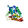 4miuC  4n9pC  4n9tC  4nmzC 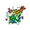 4r8nC  4rkbC  4rklC 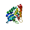 3bdcS S: Starting model for refinement C: citing same article ( |
|---|---|
| Similar structure data |


-Links
 PDBj
PDBj- Assembly
| Deposited unit | 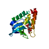
| ||||||||
|---|---|---|---|---|---|---|---|---|---|
| 1 |
| ||||||||
| Unit cell |
|
-Components
| #1: Protein | Micrococcal nuclease / TNase / Micrococcal nuclease / Staphylococcal nuclease Mass: 16119.355 Da / Num. of mol.: 1 / Fragment: Nuclease A (UNP residues 83-231) / Mutation: V23T,G50F,V51N,V66A,V99T,P117G,H124L,S128A Source method: isolated from a genetically manipulated source Source: (gene. exp.) Staphylococcus aureus (bacteria) / Gene: nuc / Plasmid: pET24a+ / Production host: Escherichia coli (E. coli) / Strain (production host): BL21(DE3) / References: UniProt: P00644, micrococcal nuclease |
|---|---|
| #2: Chemical | ChemComp-CA /   Mass: 40.078 Da / Num. of mol.: 1 / Source method: obtained synthetically / Formula: Ca Mass: 40.078 Da / Num. of mol.: 1 / Source method: obtained synthetically / Formula: Ca |
| #3: Chemical | ChemComp-THP / Thymidine diphosphate  Type: DNA linking / Mass: 402.188 Da / Num. of mol.: 1 / Source method: obtained synthetically / Formula: C10H16N2O11P2 Type: DNA linking / Mass: 402.188 Da / Num. of mol.: 1 / Source method: obtained synthetically / Formula: C10H16N2O11P2 |
| #4: Water | ChemComp-HOH / Water Mass: 18.015 Da / Num. of mol.: 132 / Source method: isolated from a natural source / Formula: H2O Mass: 18.015 Da / Num. of mol.: 132 / Source method: isolated from a natural source / Formula: H2O |
-Experimental details
-Experiment
| Experiment | Method: X-RAY DIFFRACTION / Number of used crystals: 1 |
|---|
- Sample preparation
| Crystal | Density Matthews: 2.22 Å3/Da / Density % sol: 44.57 % |
|---|---|
| Crystal grow | Temperature: 277 K / Method: vapor diffusion, hanging drop / pH: 8 Details: 20% MPD, 25 mM potassium phosphate, calcium chloride, pdTp, pH 8.0, VAPOR DIFFUSION, HANGING DROP, temperature 277K |
-Data collection
| Diffraction | Mean temperature: 100 K | |||||||||||||||||||||||||||||||||||||||||||||||||||||||||||||||||||||||||||||||||||||||||||||||||||||||||||||||||||||||||||||||||||||||||||||||||||
|---|---|---|---|---|---|---|---|---|---|---|---|---|---|---|---|---|---|---|---|---|---|---|---|---|---|---|---|---|---|---|---|---|---|---|---|---|---|---|---|---|---|---|---|---|---|---|---|---|---|---|---|---|---|---|---|---|---|---|---|---|---|---|---|---|---|---|---|---|---|---|---|---|---|---|---|---|---|---|---|---|---|---|---|---|---|---|---|---|---|---|---|---|---|---|---|---|---|---|---|---|---|---|---|---|---|---|---|---|---|---|---|---|---|---|---|---|---|---|---|---|---|---|---|---|---|---|---|---|---|---|---|---|---|---|---|---|---|---|---|---|---|---|---|---|---|---|---|---|
| Diffraction source | Source: SYNCHROTRON / Site: NSLS  / Beamline: X25 / Wavelength: 0.979 Å / Beamline: X25 / Wavelength: 0.979 Å | |||||||||||||||||||||||||||||||||||||||||||||||||||||||||||||||||||||||||||||||||||||||||||||||||||||||||||||||||||||||||||||||||||||||||||||||||||
| Detector | Type: DECTRIS PILATUS 6M / Detector: PIXEL / Date: Sep 25, 2011 Details: Meridionally-bent fused silica mirror with palladium and uncoated stripes vertically-focusing at 6.6:1 demagnification | |||||||||||||||||||||||||||||||||||||||||||||||||||||||||||||||||||||||||||||||||||||||||||||||||||||||||||||||||||||||||||||||||||||||||||||||||||
| Radiation | Monochromator: Double crystal Si(111) with cryogenically-cooled first crystal and sagittally-bent second crystal horizontally-focusing at 3.3:1 demagnification Protocol: SINGLE WAVELENGTH / Monochromatic (M) / Laue (L): M / Scattering type: x-ray | |||||||||||||||||||||||||||||||||||||||||||||||||||||||||||||||||||||||||||||||||||||||||||||||||||||||||||||||||||||||||||||||||||||||||||||||||||
| Radiation wavelength | Wavelength: 0.979 Å / Relative weight: 1 | |||||||||||||||||||||||||||||||||||||||||||||||||||||||||||||||||||||||||||||||||||||||||||||||||||||||||||||||||||||||||||||||||||||||||||||||||||
| Reflection | Resolution: 1.55→50 Å / Num. all: 20536 / Num. obs: 20536 / % possible obs: 99.2 % / Observed criterion σ(F): 0 / Observed criterion σ(I): 0 / Redundancy: 6.3 % / Biso Wilson estimate: 29.1 Å2 / Rmerge(I) obs: 0.044 / Χ2: 1.849 / Net I/σ(I): 24 | |||||||||||||||||||||||||||||||||||||||||||||||||||||||||||||||||||||||||||||||||||||||||||||||||||||||||||||||||||||||||||||||||||||||||||||||||||
| Reflection shell | Diffraction-ID: 1
|
-Phasing
| Phasing | Method: molecular replacement | |||||||||
|---|---|---|---|---|---|---|---|---|---|---|
| Phasing MR | Model details: Phaser MODE: MR_AUTO
|
- Processing
| Software |
| |||||||||||||||||||||||||||||||||||||||||||||
|---|---|---|---|---|---|---|---|---|---|---|---|---|---|---|---|---|---|---|---|---|---|---|---|---|---|---|---|---|---|---|---|---|---|---|---|---|---|---|---|---|---|---|---|---|---|---|
| Refinement | Method to determine structure: MOLECULAR REPLACEMENT Starting model: PDB ENTRY 3BDC Resolution: 1.55→38.34 Å / Cor.coef. Fo:Fc: 0.966 / Cor.coef. Fo:Fc free: 0.937 / WRfactor Rfree: 0.2536 / WRfactor Rwork: 0.1987 / Occupancy max: 1 / Occupancy min: 0.3 / FOM work R set: 0.8454 / SU B: 1.563 / SU ML: 0.058 / SU R Cruickshank DPI: 0.0846 / SU Rfree: 0.0928 / Cross valid method: THROUGHOUT / σ(F): 0 / σ(I): 0 / ESU R: 0.085 / ESU R Free: 0.093 / Stereochemistry target values: MAXIMUM LIKELIHOOD Details: HYDROGENS HAVE BEEN USED IF PRESENT IN THE INPUT U VALUES : REFINED INDIVIDUALLY
| |||||||||||||||||||||||||||||||||||||||||||||
| Solvent computation | Ion probe radii: 0.8 Å / Shrinkage radii: 0.8 Å / VDW probe radii: 1.2 Å / Solvent model: MASK | |||||||||||||||||||||||||||||||||||||||||||||
| Displacement parameters | Biso max: 67.24 Å2 / Biso mean: 23.5961 Å2 / Biso min: 11.72 Å2
| |||||||||||||||||||||||||||||||||||||||||||||
| Refinement step | Cycle: LAST / Resolution: 1.55→38.34 Å
| |||||||||||||||||||||||||||||||||||||||||||||
| Refine LS restraints |
| |||||||||||||||||||||||||||||||||||||||||||||
| LS refinement shell | Resolution: 1.55→1.59 Å / Total num. of bins used: 20
|
