 Movie
Movie Controller

[English] 日本語
 Yorodumi
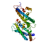
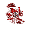
- PDB-4p1t: Crystal structure of the DBL3X-DBL4epsilon double domain from the... -
+ Open data

- Basic information
| Entry | Database: PDB / ID: 4p1t | ||||||
|---|---|---|---|---|---|---|---|
| Title | Crystal structure of the DBL3X-DBL4epsilon double domain from the extracellular part of VAR2CSA PfEMP1 from Plasmodium falciparum | ||||||
 Components Components | Erythrocyte membrane protein 1 Red blood cell Red blood cell | ||||||
 Keywords Keywords | MEMBRANE PROTEIN / PfEMP1 / CSA / DBL fold | ||||||
| Function / homology |  Function and homology information Function and homology information | ||||||
| Biological species |  Plasmodium falciparum (malaria parasite P. falciparum) Plasmodium falciparum (malaria parasite P. falciparum) | ||||||
| Method | X-RAY DIFFRACTION / SYNCHROTRON / Resolution: 2.9 Å | ||||||
 Authors Authors | Gangnard, S. / Dechavanne, S. / Srivastava, A. / Amirat, F. / Gamain, B. / Lewit-Bentley, A. / Bentley, G.A. | ||||||
 Citation Citation | Journal: Sci Rep / Year: 2015 Title: Structure of the DBL3X-DBL4 epsilon region of the VAR2CSA placental malaria vaccine candidate: insight into DBL domain interactions. Authors: Gangnard, S. / Lewit-Bentley, A. / Dechavanne, S. / Srivastava, A. / Amirat, F. / Bentley, G.A. / Gamain, B. | ||||||
| History |
|
- Structure visualization
| Structure viewer | Molecule: MolmilJmol/JSmol |
|---|
- Downloads & links
-Download
| PDBx/mmCIF format | 4p1t.cif.gz | 144.1 KB | Display | PDBx/mmCIF format |
|---|---|---|---|---|
| PDB format | pdb4p1t.ent.gz | 108.6 KB | Display | PDB format |
| PDBx/mmJSON format | 4p1t.json.gz | Tree view | PDBx/mmJSON format | |
| Others |  Other downloads Other downloads |
-Validation report
| Arichive directory | https://data.pdbj.org/pub/pdb/validation_reports/p1/4p1tftp://data.pdbj.org/pub/pdb/validation_reports/p1/4p1t | HTTPS FTP |
|---|
-Related structure data
| Related structure data | |
|---|---|
| Similar structure data |


-Links
 PDBj
PDBj- Assembly
| Deposited unit | 
| ||||||||
|---|---|---|---|---|---|---|---|---|---|
| 1 |
| ||||||||
| Unit cell |
|
-Components
| #1: Protein | Red blood cell Mass: 86439.617 Da / Num. of mol.: 1 Fragment: DBL3X-DBL4epsilon double domain (UNP residues 1215-1950) Source method: isolated from a genetically manipulated source Source: (gene. exp.) Plasmodium falciparum (malaria parasite P. falciparum)Strain: FCR-3 / Gene: var2csa / Plasmid: pET21 / Production host:  Escherichia coli (E. coli) / Strain (production host): SHuffle / References: UniProt: Q6UDW7 Escherichia coli (E. coli) / Strain (production host): SHuffle / References: UniProt: Q6UDW7 |
|---|---|
| #2: Water | ChemComp-HOH / Water Mass: 18.015 Da / Num. of mol.: 14 / Source method: isolated from a natural source / Formula: H2O Mass: 18.015 Da / Num. of mol.: 14 / Source method: isolated from a natural source / Formula: H2O |
-Experimental details
-Experiment
| Experiment | Method: X-RAY DIFFRACTION / Number of used crystals: 1 |
|---|
- Sample preparation
| Crystal | Density Matthews: 2.294 Å3/Da / Density % sol: 46.4 % |
|---|---|
| Crystal grow | Temperature: 292 K / Method: vapor diffusion, hanging drop / pH: 6.5 Details: 18% PEG 3000, 100 mM Bis-Tris buffer, 300 mM sodium chloride |
-Data collection
| Diffraction | Mean temperature: 100 K |
|---|---|
| Diffraction source | Source: SYNCHROTRON / Site: SOLEIL  / Beamline: PROXIMA 1 / Wavelength: 0.98011 Å / Beamline: PROXIMA 1 / Wavelength: 0.98011 Å |
| Detector | Type: ADSC QUANTUM 1 / Detector: CCD / Date: Mar 27, 2011 / Details: KB-MIRRORS |
| Radiation | Monochromator: CHANNEL-CUT SI (111) / Protocol: SINGLE WAVELENGTH / Scattering type: x-ray |
| Radiation wavelength | Wavelength: 0.98011 Å / Relative weight: 1 |
| Reflection | Resolution: 2.9→28.16 Å / Num. obs: 17568 / % possible obs: 92.1 % / Observed criterion σ(F): 0 / Observed criterion σ(I): 0 / Redundancy: 6.8 % / Biso Wilson estimate: 56.41 Å2 / Rmerge(I) obs: 0.263 / Rsym value: 0.243 / Net I/av σ(I): 3.1 / Net I/σ(I): 6.8 |
| Reflection shell | Resolution: 2.9→2.95 Å / Redundancy: 1.6 % / Rmerge(I) obs: 0.936 / Mean I/σ(I) obs: 0.8 / % possible all: 92.1 |
- Processing
| Software | Name: BUSTER / Version: 2.11.4 / Classification: refinement | ||||||||||||||||||||||||||||||||||||||||||||||||||||||||||||||||||||||||||||||||||||||||||||||||||||||||||||||||||
|---|---|---|---|---|---|---|---|---|---|---|---|---|---|---|---|---|---|---|---|---|---|---|---|---|---|---|---|---|---|---|---|---|---|---|---|---|---|---|---|---|---|---|---|---|---|---|---|---|---|---|---|---|---|---|---|---|---|---|---|---|---|---|---|---|---|---|---|---|---|---|---|---|---|---|---|---|---|---|---|---|---|---|---|---|---|---|---|---|---|---|---|---|---|---|---|---|---|---|---|---|---|---|---|---|---|---|---|---|---|---|---|---|---|---|---|
| Refinement | Resolution: 2.9→28.16 Å / Cor.coef. Fo:Fc: 0.879 / Cor.coef. Fo:Fc free: 0.821 / Cross valid method: THROUGHOUT / σ(F): 0 / SU Rfree Blow DPI: 0.436
| ||||||||||||||||||||||||||||||||||||||||||||||||||||||||||||||||||||||||||||||||||||||||||||||||||||||||||||||||||
| Displacement parameters | Biso mean: 60.75 Å2
| ||||||||||||||||||||||||||||||||||||||||||||||||||||||||||||||||||||||||||||||||||||||||||||||||||||||||||||||||||
| Refine analyze | Luzzati coordinate error obs: 0.361 Å | ||||||||||||||||||||||||||||||||||||||||||||||||||||||||||||||||||||||||||||||||||||||||||||||||||||||||||||||||||
| Refinement step | Cycle: final / Resolution: 2.9→28.16 Å
| ||||||||||||||||||||||||||||||||||||||||||||||||||||||||||||||||||||||||||||||||||||||||||||||||||||||||||||||||||
| Refine LS restraints |
| ||||||||||||||||||||||||||||||||||||||||||||||||||||||||||||||||||||||||||||||||||||||||||||||||||||||||||||||||||
| LS refinement shell | Resolution: 2.9→3.08 Å / Total num. of bins used: 9
|
