Group: Atomic model / Data collection ...Atomic model / Data collection / Database references / Derived calculations / Other / Refinement description / Source and taxonomy / Structure summary / Version format compliance
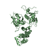
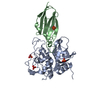
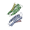
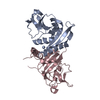
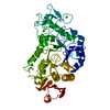
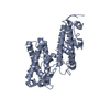
ERYTHROCYTEMEMBRANEPROTEIN1 (PFEMP1) / Red blood cell / VAR2CSA
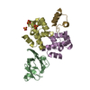
Mass: 35633.297 Da / Num. of mol.: 2 / Fragment: DBL6 EPSILON, RESIDUES 2333-2634 / Mutation: YES Source method: isolated from a genetically manipulated source Source: (gene. exp.) PLASMODIUM FALCIPARUM (malaria parasite P. falciparum) Strain: 3D7 / Description: GENOMIC DNA FROM MR4 / Cell: ERYTHROCYTE / Production host: ESCHERICHIA COLI (E. coli) / References: UniProt: Q8I639
Compound details
ENGINEERED RESIDUE IN CHAIN A, CYS 2480 TO SER ENGINEERED RESIDUE IN CHAIN B, CYS 2480 TO SER
Sequence details
THE STRUCTURE IS OF THE C2480S MUTANT.
-
Experimental details
-
Experiment
Experiment
Method: X-RAY DIFFRACTION / Number of used crystals: 1
-
Sample preparation
Crystal
Density Matthews: 2.33 Å3/Da / Density % sol: 46.8 % / Description: NONE
Crystal grow
pH: 8 / Details: pH 8
-
Data collection
Diffraction
ID
Mean temperature (K)
Crystal-ID
1
100
1
2
100
1
Diffraction source
Source
Site
Beamline
ID
Wavelength
Wavelength (Å)
SYNCHROTRON
Diamond
I02
1
0.92
SYNCHROTRON
ESRF
BM14
2
0.97855, 0.97871, 0.91999
Detector
Type
ID
Detector
Date
ADSC CCD
1
CCD
Aug 8, 2008
MARRESEARCH
2
IMAGE PLATE
Radiation
ID
Protocol
Monochromatic (M) / Laue (L)
Scattering type
Wavelength-ID
1
MAD
M
x-ray
1
2
M
x-ray
1
Radiation wavelength
ID
Wavelength (Å)
Relative weight
1
0.92
1
2
0.97855
1
3
0.97871
1
4
0.91999
1
Reflection
Resolution: 3→111.1 Å / Num. obs: 14481 / % possible obs: 99.9 % / Observed criterion σ(I): 2 / Redundancy: 5.8 % / Rmerge(I) obs: 0.1 / Net I/σ(I): 14.2
Reflection shell
Resolution: 3→3.16 Å / Redundancy: 6.2 % / Rmerge(I) obs: 0.6 / Mean I/σ(I) obs: 2.9 / % possible all: 100
-
Processing
Software
Name
Version
Classification
REFMAC
5.2.0019
refinement
MOSFLM
datareduction
SCALA
datascaling
autoSHARP
phasing
Refinement
Method to determine structure: MAD Starting model: NONE Resolution: 3→83.62 Å / Cor.coef. Fo:Fc: 0.875 / Cor.coef. Fo:Fc free: 0.855 / SU B: 60.736 / SU ML: 0.49 / Cross valid method: THROUGHOUT / ESU R Free: 0.572 Stereochemistry target values: MAXIMUM LIKELIHOOD WITH PHASES Details: HYDROGENS HAVE BEEN ADDED IN THE RIDING POSITIONS.
Rfactor
Num. reflection
% reflection
Selection details
Rfree
0.325
723
5 %
RANDOM
Rwork
0.289
-
-
-
obs
0.29
13680
99.8 %
-
Solvent computation
Ion probe radii: 0.8 Å / Shrinkage radii: 0.8 Å / VDW probe radii: 1.2 Å / Solvent model: MASK
 Movie
Movie Controller


 Open data
 Basic information
 Components
 Red blood cell
  Keywords
 Function and homology information
 Authors
 Citation
 Structure visualization
 Downloads & links
 Other downloads
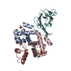
 PDBj
PDBj Assembly


 Sample preparation

 Processing