 Movie
Movie Controller

[English] 日本語
 Yorodumi
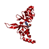
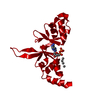
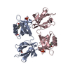
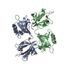
- PDB-4p1o: Crystal structure of the Bateman domain of murine magnesium trans... -
+ Open data

- Basic information
| Entry | Database: PDB / ID: 4p1o | ||||||
|---|---|---|---|---|---|---|---|
| Title | Crystal structure of the Bateman domain of murine magnesium transporter CNNM2 bound to ATP-Mg | ||||||
 Components Components | Metal transporter CNNM2 | ||||||
 Keywords Keywords |  TRANSPORT PROTEIN / magnesium homeostasis / transport / hypomagnesemia / rare diseases / ACDP / Cyclin M TRANSPORT PROTEIN / magnesium homeostasis / transport / hypomagnesemia / rare diseases / ACDP / Cyclin M | ||||||
| Function / homology |  Function and homology information Function and homology informationmagnesium ion transport / magnesium ion homeostasis / magnesium ion transmembrane transporter activity / basolateral plasma membrane / ATP binding / plasma membraneSimilarity search - Function | ||||||
| Biological species |  Mus musculus (house mouse) Mus musculus (house mouse) | ||||||
| Method | X-RAY DIFFRACTION / MOLECULAR REPLACEMENT / Resolution: 3.06 Å | ||||||
 Authors Authors | Corral-Rodriguez, M.A. / Stuiver, M. / Abascal-Palacios, G. / Diercks, T. / Oyenarte, I. / Ereno-Orbea, J. / Encinar, J.A. / Spiwok, V. / Terashima, H. / Accardi, A. ...Corral-Rodriguez, M.A. / Stuiver, M. / Abascal-Palacios, G. / Diercks, T. / Oyenarte, I. / Ereno-Orbea, J. / Encinar, J.A. / Spiwok, V. / Terashima, H. / Accardi, A. / Muller, D. / Martinez-Cruz, L.A. | ||||||
 Citation Citation | Journal: To Be Published Title: Structural and ligand binding properties of the Bateman domain of human magnesium transporters CNNM2 and CNNM4 Authors: Corral-Rodriguez, M.A. / Stuiver, M. / Abascal-Palacios, G. / Diercks, T. / Oyenarte, I. / Ereno-Orbea, J. / Encinar, J.A. / Spiwok, V. / Terashima, H. / Accardi, A. / Muller, D. / Martinez-Cruz, L.A. | ||||||
| History |
|
- Structure visualization
| Structure viewer | Molecule: MolmilJmol/JSmol |
|---|
- Downloads & links
-Download
| PDBx/mmCIF format | 4p1o.cif.gz | 138.8 KB | Display | PDBx/mmCIF format |
|---|---|---|---|---|
| PDB format | pdb4p1o.ent.gz | 110.4 KB | Display | PDB format |
| PDBx/mmJSON format | 4p1o.json.gz | Tree view | PDBx/mmJSON format | |
| Others |  Other downloads Other downloads |
-Validation report
| Arichive directory | https://data.pdbj.org/pub/pdb/validation_reports/p1/4p1oftp://data.pdbj.org/pub/pdb/validation_reports/p1/4p1o | HTTPS FTP |
|---|
-Related structure data
| Related structure data |  4iy0C  4iy2C  4iy4C  4iysC  4p1gC  4p1f 4p1p C: citing same article ( |
|---|---|
| Similar structure data |


-Links
 PDBj
PDBj
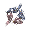
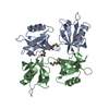
- Assembly
| Deposited unit | 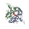
| ||||||||
|---|---|---|---|---|---|---|---|---|---|
| 1 |
| ||||||||
| Unit cell |
|
-Components
| #1: Protein | Mass: 17395.861 Da / Num. of mol.: 2 / Fragment: UNP residues 430-580 Source method: isolated from a genetically manipulated source Source: (gene. exp.) Mus musculus (house mouse) / Gene: Cnnm2, Acdp2 / Production host:  Escherichia coli (E. coli) / References: UniProt: Q3TWN3 Escherichia coli (E. coli) / References: UniProt: Q3TWN3#2: Chemical |   Mass: 24.305 Da / Num. of mol.: 2 / Source method: obtained synthetically / Formula: Mg Mass: 24.305 Da / Num. of mol.: 2 / Source method: obtained synthetically / Formula: Mg#3: Chemical | Adenosine triphosphate  Mass: 507.181 Da / Num. of mol.: 2 / Source method: obtained synthetically / Formula: C10H16N5O13P3 / Comment: ATP, energy-carrying molecule*YM Mass: 507.181 Da / Num. of mol.: 2 / Source method: obtained synthetically / Formula: C10H16N5O13P3 / Comment: ATP, energy-carrying molecule*YM |
|---|
-Experimental details
-Experiment
| Experiment | Method: X-RAY DIFFRACTION / Number of used crystals: 1 |
|---|
- Sample preparation
| Crystal | Density Matthews: 3.93 Å3/Da / Density % sol: 68.71 % |
|---|---|
| Crystal grow | Temperature: 293 K / Method: vapor diffusion, hanging drop Details: 0.1M Sodium Acetate, 3.5M Ammonium Acetate, 5mM Magnesium chloride, 10mM ATP |
-Data collection
| Diffraction | Mean temperature: 100 K |
|---|---|
| Diffraction source | Source: ROTATING ANODE / Type: BRUKER AXS MICROSTAR-H / Wavelength: 1.54179 Å |
| Detector | Type: MAR scanner 345 mm plate / Detector: IMAGE PLATE / Date: Dec 11, 2013 |
| Radiation | Protocol: SINGLE WAVELENGTH / Scattering type: x-ray |
| Radiation wavelength | Wavelength: 1.54179 Å / Relative weight: 1 |
| Reflection | Resolution: 3.06→33.08 Å / Num. obs: 10982 / % possible obs: 96.04 % / Redundancy: 3.8 % / Biso Wilson estimate: 65.79 Å2 / Net I/σ(I): 12.43 |
- Processing
| Software |
| |||||||||||||||||||||||||||||||||||||||||||||||||||||||||||||||
|---|---|---|---|---|---|---|---|---|---|---|---|---|---|---|---|---|---|---|---|---|---|---|---|---|---|---|---|---|---|---|---|---|---|---|---|---|---|---|---|---|---|---|---|---|---|---|---|---|---|---|---|---|---|---|---|---|---|---|---|---|---|---|---|---|
| Refinement | Method to determine structure: MOLECULAR REPLACEMENT / Resolution: 3.06→33.08 Å / FOM work R set: 0.7956 / SU ML: 0.34 / Cross valid method: FREE R-VALUE / σ(F): 1.35 / Phase error: 26.33 / Stereochemistry target values: ML
| |||||||||||||||||||||||||||||||||||||||||||||||||||||||||||||||
| Solvent computation | Shrinkage radii: 0.9 Å / VDW probe radii: 1.11 Å / Solvent model: FLAT BULK SOLVENT MODEL | |||||||||||||||||||||||||||||||||||||||||||||||||||||||||||||||
| Displacement parameters | Biso max: 189.76 Å2 / Biso mean: 73.71 Å2 / Biso min: 38.54 Å2 | |||||||||||||||||||||||||||||||||||||||||||||||||||||||||||||||
| Refinement step | Cycle: final / Resolution: 3.06→33.08 Å
| |||||||||||||||||||||||||||||||||||||||||||||||||||||||||||||||
| Refine LS restraints |
| |||||||||||||||||||||||||||||||||||||||||||||||||||||||||||||||
| LS refinement shell | Refine-ID: X-RAY DIFFRACTION / Total num. of bins used: 8
| |||||||||||||||||||||||||||||||||||||||||||||||||||||||||||||||
| Refinement TLS params. | Method: refined / Origin x: 111.0417 Å / Origin y: 45.0052 Å / Origin z: -0.2612 Å
| |||||||||||||||||||||||||||||||||||||||||||||||||||||||||||||||
| Refinement TLS group |
|
