 Movie
Movie Controller

[English] 日本語
 Yorodumi
- PDB-1obz: Crystal structure of the complex of the PDZ tandem of syntenin wi... -
+ Open data

- Basic information
| Entry | Database: PDB / ID: 1obz | ||||||
|---|---|---|---|---|---|---|---|
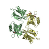
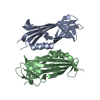
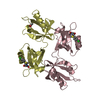
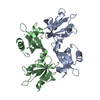
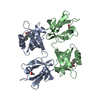
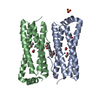
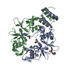
| Title | Crystal structure of the complex of the PDZ tandem of syntenin with an interleukin 5 receptor alpha peptide. | ||||||
 Components Components |
| ||||||
 Keywords Keywords |  CELL ADHESION / ADHESION-COMPLEX / PDZ DOMAIN / SIGNAL TRANSDUCTION / NUCLEAR PROTEIN CELL ADHESION / ADHESION-COMPLEX / PDZ DOMAIN / SIGNAL TRANSDUCTION / NUCLEAR PROTEIN | ||||||
| Function / homology |  Function and homology informationinterleukin-5 receptor activity / regulation of interleukin-5 production / interleukin-5 receptor complex / interleukin-5 receptor binding / positive regulation of extracellular exosome assembly / syndecan binding / Neurofascin interactions / presynapse assembly / neurexin family protein binding / cytoskeletal anchor activity ...interleukin-5 receptor activity / regulation of interleukin-5 production / interleukin-5 receptor complex / interleukin-5 receptor binding / positive regulation of extracellular exosome assembly / syndecan binding / Neurofascin interactions / presynapse assembly / neurexin family protein binding / cytoskeletal anchor activity / substrate-dependent cell migration, cell extension / interleukin-5-mediated signaling pathway / positive regulation of exosomal secretion / positive regulation of leukocyte proliferation / negative regulation of receptor internalization / frizzled binding / inflammatory response to antigenic stimulus / protein targeting to membrane / Ephrin signaling / cytokine receptor activity / RIPK1-mediated regulated necrosis / cytokine binding / positive regulation of transforming growth factor beta receptor signaling pathway / growth factor binding / Interleukin-3, Interleukin-5 and GM-CSF signaling / Interleukin receptor SHC signaling / positive regulation of epithelial to mesenchymal transition / positive regulation of phosphorylation / cell adhesion molecule binding / regulation of mitotic cell cycle / ionotropic glutamate receptor binding / phosphatidylinositol-4,5-bisphosphate binding / protein sequestering activity / ephrin receptor binding / positive regulation of JNK cascade / adherens junction / Regulation of necroptotic cell death / cytokine-mediated signaling pathway / azurophil granule lumen / extracellular vesicle / melanosome / presynapse / actin cytoskeleton organization / RAF/MAP kinase cascade / positive regulation of cell growth / chemical synaptic transmission / nuclear membrane / Ras protein signal transduction / blood microparticle / cytoskeleton / receptor complex / intracellular signal transduction / positive regulation of cell migration / membrane raft / protein heterodimerization activity / external side of plasma membrane / focal adhesion / Neutrophil degranulation / positive regulation of cell population proliferation / protein-containing complex binding / endoplasmic reticulum membrane / negative regulation of transcription by RNA polymerase II / signal transduction / extracellular space / extracellular exosome / extracellular region / nucleoplasm / membrane / identical protein binding / nucleus / plasma membrane / cytosol / cytoplasm Function and homology informationinterleukin-5 receptor activity / regulation of interleukin-5 production / interleukin-5 receptor complex / interleukin-5 receptor binding / positive regulation of extracellular exosome assembly / syndecan binding / Neurofascin interactions / presynapse assembly / neurexin family protein binding / cytoskeletal anchor activity ...interleukin-5 receptor activity / regulation of interleukin-5 production / interleukin-5 receptor complex / interleukin-5 receptor binding / positive regulation of extracellular exosome assembly / syndecan binding / Neurofascin interactions / presynapse assembly / neurexin family protein binding / cytoskeletal anchor activity / substrate-dependent cell migration, cell extension / interleukin-5-mediated signaling pathway / positive regulation of exosomal secretion / positive regulation of leukocyte proliferation / negative regulation of receptor internalization / frizzled binding / inflammatory response to antigenic stimulus / protein targeting to membrane / Ephrin signaling / cytokine receptor activity / RIPK1-mediated regulated necrosis / cytokine binding / positive regulation of transforming growth factor beta receptor signaling pathway / growth factor binding / Interleukin-3, Interleukin-5 and GM-CSF signaling / Interleukin receptor SHC signaling / positive regulation of epithelial to mesenchymal transition / positive regulation of phosphorylation / cell adhesion molecule binding / regulation of mitotic cell cycle / ionotropic glutamate receptor binding / phosphatidylinositol-4,5-bisphosphate binding / protein sequestering activity / ephrin receptor binding / positive regulation of JNK cascade / adherens junction / Regulation of necroptotic cell death / cytokine-mediated signaling pathway / azurophil granule lumen / extracellular vesicle / melanosome / presynapse / actin cytoskeleton organization / RAF/MAP kinase cascade / positive regulation of cell growth / chemical synaptic transmission / nuclear membrane / Ras protein signal transduction / blood microparticle / cytoskeleton / receptor complex / intracellular signal transduction / positive regulation of cell migration / membrane raft / protein heterodimerization activity / external side of plasma membrane / focal adhesion / Neutrophil degranulation / positive regulation of cell population proliferation / protein-containing complex binding / endoplasmic reticulum membrane / negative regulation of transcription by RNA polymerase II / signal transduction / extracellular space / extracellular exosome / extracellular region / nucleoplasm / membrane / identical protein binding / nucleus / plasma membrane / cytosol / cytoplasmSimilarity search - Function | ||||||
| Biological species |  HOMO SAPIENS (human) HOMO SAPIENS (human) | ||||||
| Method | X-RAY DIFFRACTION / SYNCHROTRON / MOLECULAR REPLACEMENT / Resolution: 1.7 Å | ||||||
 Authors Authors | Kang, B.S. / Cooper, D.R. / Devedjiev, Y. / Derewenda, U. / Derewenda, Z.S. | ||||||
 Citation Citation | Journal: Structure / Year: 2003 Title: Molecular Roots of Degenerate Specificity in Syntenin'S Pdz2 Domain: Reassessment of the Pdz Recognition Paradigm Authors: Kang, B.S. / Cooper, D.R. / Devedjiev, Y. / Derewenda, U. / Derewenda, Z.S. | ||||||
| History |
| ||||||
| Remark 700 | SHEET THE SHEET STRUCTURE OF THIS MOLECULE IS BIFURCATED. IN ORDER TO REPRESENT THIS FEATURE IN ... SHEET THE SHEET STRUCTURE OF THIS MOLECULE IS BIFURCATED. IN ORDER TO REPRESENT THIS FEATURE IN THE SHEET RECORDS BELOW, TWO SHEETS ARE DEFINED. |
- Structure visualization
| Structure viewer | Molecule: MolmilJmol/JSmol |
|---|
- Downloads & links
-Download
| PDBx/mmCIF format | 1obz.cif.gz | 147.8 KB | Display | PDBx/mmCIF format |
|---|---|---|---|---|
| PDB format | pdb1obz.ent.gz | 115.7 KB | Display | PDB format |
| PDBx/mmJSON format | 1obz.json.gz | Tree view | PDBx/mmJSON format | |
| Others |  Other downloads Other downloads |
-Validation report
| Arichive directory | https://data.pdbj.org/pub/pdb/validation_reports/ob/1obzftp://data.pdbj.org/pub/pdb/validation_reports/ob/1obz | HTTPS FTP |
|---|
-Related structure data
| Related structure data |  1nteC  1obxC  1obyC  1n99S C: citing same article ( S: Starting model for refinement |
|---|---|
| Similar structure data |
-Links
 PDBj
PDBj


- Assembly
| Deposited unit | 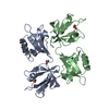
| |||||||||
|---|---|---|---|---|---|---|---|---|---|---|
| 1 |
| |||||||||
| Unit cell |
| |||||||||
| Components on special symmetry positions |
| |||||||||
| Details | THE TRIMER IS FORMED BY THE COMPLEX OF CHAINS A AND BWITH ONE MOLECULE OF CHAIN P |
-Components
| #1: Protein | Mass: 18018.688 Da / Num. of mol.: 2 / Fragment: PDZ2, RESIDUES 113-273 Source method: isolated from a genetically manipulated source Source: (gene. exp.) HOMO SAPIENS (human) / Production host:  ESCHERICHIA COLI (E. coli) / Strain (production host): BL21 / References: UniProt: O00560 ESCHERICHIA COLI (E. coli) / Strain (production host): BL21 / References: UniProt: O00560#2: Protein/peptide | | Mass: 938.975 Da / Num. of mol.: 1 / Fragment: LAST 8 RESIDUES, RESIDUES 413-420 / Source method: obtained synthetically / Source: (synth.) HOMO SAPIENS (human) / References: UniProt: Q01344#3: Chemical | ChemComp-ACT / Acetate  Mass: 59.044 Da / Num. of mol.: 4 / Source method: obtained synthetically / Formula: C2H3O2 Mass: 59.044 Da / Num. of mol.: 4 / Source method: obtained synthetically / Formula: C2H3O2#4: Water | ChemComp-HOH / | Water Mass: 18.015 Da / Num. of mol.: 658 / Source method: isolated from a natural source / Formula: H2O Mass: 18.015 Da / Num. of mol.: 658 / Source method: isolated from a natural source / Formula: H2OCompound details | MOL_ID 1: FUNCTIONS AS AN ADAPTER PROTEIN. MAY ALSO FUNCTION TO COUPLE SYNDECANS TO CYTOSKELETAL ...MOL_ID 1: FUNCTIONS AS AN ADAPTER PROTEIN. MAY ALSO FUNCTION TO COUPLE SYNDECANS TO CYTOSKELET | |
|---|
-Experimental details
-Experiment
| Experiment | Method: X-RAY DIFFRACTION / Number of used crystals: 1 |
|---|
- Sample preparation
| Crystal | Density Matthews: 2.2 Å3/Da / Density % sol: 43.7 % |
|---|---|
| Crystal grow | Method: vapor diffusion, sitting drop / pH: 4.6 Details: SITTING DROP WITH 0.2 M SODIUM ACETATE, PH 4.6 28% PEG4000, 0.2 M AMMONIUM ACETATE |
-Data collection
| Diffraction | Mean temperature: 100 K |
|---|---|
| Diffraction source | Source: SYNCHROTRON / Site: NSLS  / Beamline: X9B / Wavelength: 0.97946 / Beamline: X9B / Wavelength: 0.97946 |
| Detector | Type: ADSC CCD / Detector: CCD / Date: Jan 20, 2002 |
| Radiation | Protocol: SINGLE WAVELENGTH / Monochromatic (M) / Laue (L): M / Scattering type: x-ray |
| Radiation wavelength | Wavelength: 0.97946 Å / Relative weight: 1 |
| Reflection | Resolution: 1.7→50 Å / Num. obs: 112090 / % possible obs: 95.7 % / Redundancy: 3.4 % / Rmerge(I) obs: 0.044 / Net I/σ(I): 25.4 |
| Reflection shell | Resolution: 1.7→1.76 Å / Rmerge(I) obs: 0.287 / Mean I/σ(I) obs: 3.13 / % possible all: 72.4 |
| Reflection | *PLUS Highest resolution: 1.7 Å / Lowest resolution: 50 Å / Redundancy: 3.4 % / Rmerge(I) obs: 0.044 |
| Reflection shell | *PLUS Rmerge(I) obs: 0.287 / Mean I/σ(I) obs: 3.13 |
- Processing
| Software |
| ||||||||||||||||||||||||||||||||||||||||||||||||||||||||||||||||||||||||||||||||||||||||||||||||||||||||||||||||||||||||||||||||||||||||||||||||||||||||||||||||||||||||||||||||||||||
|---|---|---|---|---|---|---|---|---|---|---|---|---|---|---|---|---|---|---|---|---|---|---|---|---|---|---|---|---|---|---|---|---|---|---|---|---|---|---|---|---|---|---|---|---|---|---|---|---|---|---|---|---|---|---|---|---|---|---|---|---|---|---|---|---|---|---|---|---|---|---|---|---|---|---|---|---|---|---|---|---|---|---|---|---|---|---|---|---|---|---|---|---|---|---|---|---|---|---|---|---|---|---|---|---|---|---|---|---|---|---|---|---|---|---|---|---|---|---|---|---|---|---|---|---|---|---|---|---|---|---|---|---|---|---|---|---|---|---|---|---|---|---|---|---|---|---|---|---|---|---|---|---|---|---|---|---|---|---|---|---|---|---|---|---|---|---|---|---|---|---|---|---|---|---|---|---|---|---|---|---|---|---|---|
| Refinement | Method to determine structure: MOLECULAR REPLACEMENT Starting model: PDB ENTRY 1N99 Resolution: 1.7→64.55 Å / Cor.coef. Fo:Fc: 0.965 / Cor.coef. Fo:Fc free: 0.93 / SU B: 2.207 / SU ML: 0.073 / Cross valid method: THROUGHOUT / ESU R: 0.172 / ESU R Free: 0.117 / Stereochemistry target values: MAXIMUM LIKELIHOOD / Details: HYDROGENS HAVE BEEN ADDED IN THE RIDING POSITIONS
| ||||||||||||||||||||||||||||||||||||||||||||||||||||||||||||||||||||||||||||||||||||||||||||||||||||||||||||||||||||||||||||||||||||||||||||||||||||||||||||||||||||||||||||||||||||||
| Solvent computation | Ion probe radii: 0.8 Å / Shrinkage radii: 0.8 Å / VDW probe radii: 1.4 Å / Solvent model: BABINET MODEL PLUS MASK | ||||||||||||||||||||||||||||||||||||||||||||||||||||||||||||||||||||||||||||||||||||||||||||||||||||||||||||||||||||||||||||||||||||||||||||||||||||||||||||||||||||||||||||||||||||||
| Displacement parameters | Biso mean: 12.64 Å2
| ||||||||||||||||||||||||||||||||||||||||||||||||||||||||||||||||||||||||||||||||||||||||||||||||||||||||||||||||||||||||||||||||||||||||||||||||||||||||||||||||||||||||||||||||||||||
| Refinement step | Cycle: LAST / Resolution: 1.7→64.55 Å
| ||||||||||||||||||||||||||||||||||||||||||||||||||||||||||||||||||||||||||||||||||||||||||||||||||||||||||||||||||||||||||||||||||||||||||||||||||||||||||||||||||||||||||||||||||||||
| Refine LS restraints |
|