 Movie
Movie Controller

[English] 日本語
 Yorodumi
- PDB-4p06: Bacterial arylsulfate sulfotransferase (ASST) H436N mutant with 4... -
+ Open data

- Basic information
| Entry | Database: PDB / ID: 4p06 | |||||||||
|---|---|---|---|---|---|---|---|---|---|---|
| Title | Bacterial arylsulfate sulfotransferase (ASST) H436N mutant with 4-methylumbelliferyl sulfate (MUS) in the active site | |||||||||
 Components Components | Arylsulfate sulfotransferase AssT | |||||||||
 Keywords Keywords |  TRANSFERASE / sulfotransferase / beta propeller / active site mutant TRANSFERASE / sulfotransferase / beta propeller / active site mutant | |||||||||
| Function / homology |  Function and homology informationaryl-sulfate sulfotransferase / arylsulfate sulfotransferase activity / aryl sulfotransferase activity / periplasmic space Function and homology informationaryl-sulfate sulfotransferase / arylsulfate sulfotransferase activity / aryl sulfotransferase activity / periplasmic spaceSimilarity search - Function | |||||||||
| Biological species |  Escherichia coli CFT073 (bacteria) Escherichia coli CFT073 (bacteria) | |||||||||
| Method | X-RAY DIFFRACTION / SYNCHROTRON / MOLECULAR REPLACEMENT / Resolution: 2.1 Å | |||||||||
 Authors Authors | Malojcic, G. / Owen, R.L. / Glockshuber, R. | |||||||||
| Funding support |  Switzerland, 1items Switzerland, 1items
| |||||||||
 Citation Citation | Journal: Biochemistry / Year: 2014 Title: Structural and mechanistic insights into the PAPS-independent sulfotransfer catalyzed by bacterial aryl sulfotransferase and the role of the DsbL/Dsbl system in its folding. Authors: Malojcic, G. / Owen, R.L. / Glockshuber, R. | |||||||||
| History |
|
- Structure visualization
| Structure viewer | Molecule: MolmilJmol/JSmol |
|---|
- Downloads & links
-Download
| PDBx/mmCIF format | 4p06.cif.gz | 230.1 KB | Display | PDBx/mmCIF format |
|---|---|---|---|---|
| PDB format | pdb4p06.ent.gz | 183.2 KB | Display | PDB format |
| PDBx/mmJSON format | 4p06.json.gz | Tree view | PDBx/mmJSON format | |
| Others |  Other downloads Other downloads |
-Validation report
| Arichive directory | https://data.pdbj.org/pub/pdb/validation_reports/p0/4p06ftp://data.pdbj.org/pub/pdb/validation_reports/p0/4p06 | HTTPS FTP |
|---|
-Related structure data
| Related structure data |  4p04C  4p05C  4p07C 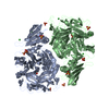 3elqS S: Starting model for refinement C: citing same article ( |
|---|---|
| Similar structure data |


-Links
 PDBj
PDBj
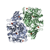
- Assembly
| Deposited unit | 
| ||||||||
|---|---|---|---|---|---|---|---|---|---|
| 1 |
| ||||||||
| 2 | 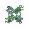
| ||||||||
| Unit cell |
| ||||||||
| Details | Gel filtration confirms the dimerization of the protein in solution |
-Components
| #1: Protein | Mass: 63822.594 Da / Num. of mol.: 2 / Mutation: H463N Source method: isolated from a genetically manipulated source Source: (gene. exp.) Escherichia coli CFT073 (bacteria) / Gene: assT, LF82_506 / Production host: Escherichia coli (E. coli)References: UniProt: E2QE64, UniProt: A0A0D6H805*PLUS, aryl-sulfate sulfotransferase#2: Chemical | Sulfate  Mass: 96.063 Da / Num. of mol.: 3 / Source method: obtained synthetically / Formula: SO4 Mass: 96.063 Da / Num. of mol.: 3 / Source method: obtained synthetically / Formula: SO4#3: Chemical |   Mass: 256.232 Da / Num. of mol.: 2 / Source method: obtained synthetically / Formula: C10H8O6S Mass: 256.232 Da / Num. of mol.: 2 / Source method: obtained synthetically / Formula: C10H8O6S#4: Water | ChemComp-HOH / | Water Mass: 18.015 Da / Num. of mol.: 16 / Source method: isolated from a natural source / Formula: H2O Mass: 18.015 Da / Num. of mol.: 16 / Source method: isolated from a natural source / Formula: H2O |
|---|
-Experimental details
-Experiment
| Experiment | Method: X-RAY DIFFRACTION |
|---|
- Sample preparation
| Crystal | Density Matthews: 3.74 Å3/Da / Density % sol: 67.12 % |
|---|---|
| Crystal grow | Temperature: 293 K / Method: vapor diffusion, sitting drop Details: sitting drop vapor diffusion method by equilibrating 1.5 ?l of protein solution (22 mg/ml, in 20 mM 4-morpholinepropanesulfonic acid/NaOH pH 7.5, 100 mM NaCl) with 0.5 ?l of reservoir ...Details: sitting drop vapor diffusion method by equilibrating 1.5 ?l of protein solution (22 mg/ml, in 20 mM 4-morpholinepropanesulfonic acid/NaOH pH 7.5, 100 mM NaCl) with 0.5 ?l of reservoir solution consisting of 1.8 M Li2SO4 and 100 mM cacodylic acid/NaOH pH 6.5. |
-Data collection
| Diffraction | Mean temperature: 100 K |
|---|---|
| Diffraction source | Source: SYNCHROTRON / Site: SLS / Beamline: X10SA / Wavelength: 1 Å |
| Detector | Type: DECTRIS PILATUS 6M / Detector: PIXEL / Date: Jul 23, 2012 |
| Radiation | Monochromator: Si(111) / Protocol: SINGLE WAVELENGTH / Monochromatic (M) / Laue (L): M / Scattering type: x-ray |
| Radiation wavelength | Wavelength: 1 Å / Relative weight: 1 |
| Reflection | Resolution: 2.1→52 Å / Num. obs: 109496 / % possible obs: 99.9 % / Redundancy: 4.5 % / Rmerge(I) obs: 0.065 / Rsym value: 0.028 / Net I/σ(I): 13.9 |
| Reflection shell | Resolution: 2.1→2.21 Å / Redundancy: 4.5 % / Rmerge(I) obs: 0.84 / Mean I/σ(I) obs: 1.8 / % possible all: 99.9 |
- Processing
| Software |
| |||||||||||||||||||||||||||||||||||||||||||||||||||||||||||||||||||||||||||||||||||||||||||||||||||||||||||||||||||||||||||||||||||||||||||||||||||
|---|---|---|---|---|---|---|---|---|---|---|---|---|---|---|---|---|---|---|---|---|---|---|---|---|---|---|---|---|---|---|---|---|---|---|---|---|---|---|---|---|---|---|---|---|---|---|---|---|---|---|---|---|---|---|---|---|---|---|---|---|---|---|---|---|---|---|---|---|---|---|---|---|---|---|---|---|---|---|---|---|---|---|---|---|---|---|---|---|---|---|---|---|---|---|---|---|---|---|---|---|---|---|---|---|---|---|---|---|---|---|---|---|---|---|---|---|---|---|---|---|---|---|---|---|---|---|---|---|---|---|---|---|---|---|---|---|---|---|---|---|---|---|---|---|---|---|---|---|
| Refinement | Method to determine structure: MOLECULAR REPLACEMENT Starting model: 3ELQ Resolution: 2.1→51.133 Å / Cross valid method: FREE R-VALUE / σ(F): 1.33 / Phase error: 26.88 / Stereochemistry target values: TWIN_LSQ_F
| |||||||||||||||||||||||||||||||||||||||||||||||||||||||||||||||||||||||||||||||||||||||||||||||||||||||||||||||||||||||||||||||||||||||||||||||||||
| Solvent computation | Shrinkage radii: 0.9 Å / VDW probe radii: 1.11 Å / Solvent model: FLAT BULK SOLVENT MODEL | |||||||||||||||||||||||||||||||||||||||||||||||||||||||||||||||||||||||||||||||||||||||||||||||||||||||||||||||||||||||||||||||||||||||||||||||||||
| Refinement step | Cycle: LAST / Resolution: 2.1→51.133 Å
| |||||||||||||||||||||||||||||||||||||||||||||||||||||||||||||||||||||||||||||||||||||||||||||||||||||||||||||||||||||||||||||||||||||||||||||||||||
| Refine LS restraints |
| |||||||||||||||||||||||||||||||||||||||||||||||||||||||||||||||||||||||||||||||||||||||||||||||||||||||||||||||||||||||||||||||||||||||||||||||||||
| LS refinement shell |
|
