 Movie
Movie Controller

[English] 日本語
 Yorodumi
- PDB-4ov0: Structure of Bacteriorhdopsin Transferred from Amphipol A8-35 to ... -
+ Open data

- Basic information
| Entry | Database: PDB / ID: 4ov0 | ||||||
|---|---|---|---|---|---|---|---|
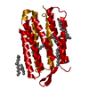
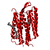
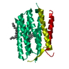
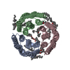
| Title | Structure of Bacteriorhdopsin Transferred from Amphipol A8-35 to a Lipidic Mesophase | ||||||
 Components Components | Bacteriorhodopsin | ||||||
 Keywords Keywords | TRANSPORT PROTEIN / Light-driven proton pump / Retinal attached via Schiff base / Membrane | ||||||
| Function / homology |  Function and homology informationphotoreceptor activity / phototransduction / proton transmembrane transport / monoatomic ion channel activity / plasma membrane Function and homology informationphotoreceptor activity / phototransduction / proton transmembrane transport / monoatomic ion channel activity / plasma membraneSimilarity search - Function | ||||||
| Biological species |  Halobacterium sp. (Halophile) Halobacterium sp. (Halophile) | ||||||
| Method | X-RAY DIFFRACTION / SYNCHROTRON / MOLECULAR REPLACEMENT / Resolution: 2 Å | ||||||
 Authors Authors | Polovinkin, V. / Gushchin, I. / Sintsov, M. / Round, E. / Balandin, T. / Chervakov, P. / Schevchenko, V. / Utrobin, P. / Popov, A. / Borshchevskiy, V. ...Polovinkin, V. / Gushchin, I. / Sintsov, M. / Round, E. / Balandin, T. / Chervakov, P. / Schevchenko, V. / Utrobin, P. / Popov, A. / Borshchevskiy, V. / Mishin, A. / Kuklin, A. / Willbold, D. / Popot, J.L. / Gordeliy, V. | ||||||
 Citation Citation | Journal: J.Membr.Biol. / Year: 2014 Title: High-resolution structure of a membrane protein transferred from amphipol to a lipidic mesophase. Authors: Polovinkin, V. / Gushchin, I. / Sintsov, M. / Round, E. / Balandin, T. / Chervakov, P. / Schevchenko, V. / Utrobin, P. / Popov, A. / Borshchevskiy, V. / Mishin, A. / Kuklin, A. / Willbold, D. ...Authors: Polovinkin, V. / Gushchin, I. / Sintsov, M. / Round, E. / Balandin, T. / Chervakov, P. / Schevchenko, V. / Utrobin, P. / Popov, A. / Borshchevskiy, V. / Mishin, A. / Kuklin, A. / Willbold, D. / Chupin, V. / Popot, J.L. / Gordeliy, V. | ||||||
| History |
|
- Structure visualization
| Structure viewer | Molecule: MolmilJmol/JSmol |
|---|
- Downloads & links
-Download
| PDBx/mmCIF format | 4ov0.cif.gz | 50 KB | Display | PDBx/mmCIF format |
|---|---|---|---|---|
| PDB format | pdb4ov0.ent.gz | 39.4 KB | Display | PDB format |
| PDBx/mmJSON format | 4ov0.json.gz | Tree view | PDBx/mmJSON format | |
| Others |  Other downloads Other downloads |
-Validation report
| Arichive directory | https://data.pdbj.org/pub/pdb/validation_reports/ov/4ov0ftp://data.pdbj.org/pub/pdb/validation_reports/ov/4ov0 | HTTPS FTP |
|---|
-Related structure data
| Similar structure data |
|---|


-Links
 PDBj
PDBj


- Assembly
| Deposited unit | 
| ||||||||
|---|---|---|---|---|---|---|---|---|---|
| 1 | 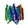
| ||||||||
| Unit cell |
|
-Components
| #1: Protein | / BR / Bacterioopsin / BO Mass: 26929.500 Da / Num. of mol.: 1 / Source method: isolated from a natural source / Source: (natural) Halobacterium sp. (Halophile) / Strain: ATCC 700922 / JCM 11081 / NRC-1 / References: UniProt: P02945 |
|---|---|
| #2: Chemical | ChemComp-RET / Retinal  Mass: 284.436 Da / Num. of mol.: 1 / Source method: obtained synthetically / Formula: C20H28O Mass: 284.436 Da / Num. of mol.: 1 / Source method: obtained synthetically / Formula: C20H28O |
| #3: Water | ChemComp-HOH / Water Mass: 18.015 Da / Num. of mol.: 14 / Source method: isolated from a natural source / Formula: H2O Mass: 18.015 Da / Num. of mol.: 14 / Source method: isolated from a natural source / Formula: H2O |
-Experimental details
-Experiment
| Experiment | Method: X-RAY DIFFRACTION / Number of used crystals: 1 |
|---|
- Sample preparation
| Crystal | Density Matthews: 2.16 Å3/Da / Density % sol: 42.99 % |
|---|---|
| Crystal grow | Temperature: 295 K / Method: vapor diffusion, hanging drop / pH: 5.6 Details: Monooleoyl, amphipol A8-35, 1-2M sodium/potassium phosphate, pH 5.6, VAPOR DIFFUSION, HANGING DROP, temperature 295K |
-Data collection
| Diffraction | Mean temperature: 100 K |
|---|---|
| Diffraction source | Source: SYNCHROTRON / Site: ESRF  / Beamline: ID23-1 / Wavelength: 0.972 Å / Beamline: ID23-1 / Wavelength: 0.972 Å |
| Detector | Type: PSI PILATUS 6M / Detector: PIXEL / Date: May 3, 2013 |
| Radiation | Protocol: SINGLE WAVELENGTH / Monochromatic (M) / Laue (L): M / Scattering type: x-ray |
| Radiation wavelength | Wavelength: 0.972 Å / Relative weight: 1 |
| Reflection | Resolution: 2→52.83 Å / Num. all: 15434 / Num. obs: 15418 / % possible obs: 99.9 % / Observed criterion σ(F): 4 / Observed criterion σ(I): 2 / Redundancy: 6.4 % / Rmerge(I) obs: 0.121 / Net I/σ(I): 9.7 |
- Processing
| Software |
| ||||||||||||||||||||||||||||||||||||||||||
|---|---|---|---|---|---|---|---|---|---|---|---|---|---|---|---|---|---|---|---|---|---|---|---|---|---|---|---|---|---|---|---|---|---|---|---|---|---|---|---|---|---|---|---|
| Refinement | Method to determine structure: MOLECULAR REPLACEMENT / Resolution: 2→52.826 Å / σ(F): 1.41 / Phase error: 22.01 / Stereochemistry target values: TWIN_LSQ_F
| ||||||||||||||||||||||||||||||||||||||||||
| Solvent computation | Shrinkage radii: 0.9 Å / VDW probe radii: 1.11 Å / Solvent model: FLAT BULK SOLVENT MODEL | ||||||||||||||||||||||||||||||||||||||||||
| Refinement step | Cycle: LAST / Resolution: 2→52.826 Å
| ||||||||||||||||||||||||||||||||||||||||||
| Refine LS restraints |
| ||||||||||||||||||||||||||||||||||||||||||
| LS refinement shell |
|
