 Movie
Movie Controller

[English] 日本語
 Yorodumi
- PDB-4ogx: Crystal structure of Fab DX-2930 in complex with human plasma kal... -
+ Open data

- Basic information
| Entry | Database: PDB / ID: 4ogx | ||||||
|---|---|---|---|---|---|---|---|
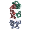
| Title | Crystal structure of Fab DX-2930 in complex with human plasma kallikrein at 2.4 Angstrom resolution | ||||||
 Components Components |
| ||||||
 Keywords Keywords | HYDROLASE/ANTIBODY /  FAB / ANTIBODY / KALLIKREIN / BLOOD / PLASMA / PLASMA KALLIKREIN- MEDIATED EDEMA / ACUTE HEREDITARY ANGIOEDEMA / HAE / HMWK / serpin C1-inhibitor / C1-INH / hereditary angioedema / HAW / bradykinin / Fletcher factor / Kininogenin / serine protease / edema / HYDROLASE-ANTIBODY complex FAB / ANTIBODY / KALLIKREIN / BLOOD / PLASMA / PLASMA KALLIKREIN- MEDIATED EDEMA / ACUTE HEREDITARY ANGIOEDEMA / HAE / HMWK / serpin C1-inhibitor / C1-INH / hereditary angioedema / HAW / bradykinin / Fletcher factor / Kininogenin / serine protease / edema / HYDROLASE-ANTIBODY complex | ||||||
| Function / homology |  Function and homology informationplasma kallikrein / Factor XII activation / Defective SERPING1 causes hereditary angioedema / positive regulation of fibrinolysis / zymogen activation / immunoglobulin complex / plasminogen activation / Defective factor XII causes hereditary angioedema / Activation of Matrix Metalloproteinases / fibrinolysis ...plasma kallikrein / Factor XII activation / Defective SERPING1 causes hereditary angioedema / positive regulation of fibrinolysis / zymogen activation / immunoglobulin complex / plasminogen activation / Defective factor XII causes hereditary angioedema / Activation of Matrix Metalloproteinases / fibrinolysis / Intrinsic Pathway of Fibrin Clot Formation / blood coagulation / adaptive immune response / immune response / serine-type endopeptidase activity / proteolysis / extracellular space / extracellular exosome / extracellular region / plasma membrane Function and homology informationplasma kallikrein / Factor XII activation / Defective SERPING1 causes hereditary angioedema / positive regulation of fibrinolysis / zymogen activation / immunoglobulin complex / plasminogen activation / Defective factor XII causes hereditary angioedema / Activation of Matrix Metalloproteinases / fibrinolysis ...plasma kallikrein / Factor XII activation / Defective SERPING1 causes hereditary angioedema / positive regulation of fibrinolysis / zymogen activation / immunoglobulin complex / plasminogen activation / Defective factor XII causes hereditary angioedema / Activation of Matrix Metalloproteinases / fibrinolysis / Intrinsic Pathway of Fibrin Clot Formation / blood coagulation / adaptive immune response / immune response / serine-type endopeptidase activity / proteolysis / extracellular space / extracellular exosome / extracellular region / plasma membraneSimilarity search - Function | ||||||
| Biological species |  Homo sapiens (human) Homo sapiens (human) | ||||||
| Method | X-RAY DIFFRACTION / MOLECULAR REPLACEMENT / molecular replacement / Resolution: 2.4 Å | ||||||
 Authors Authors | Edwards, T.E. / Clifton, M.C. / Abendroth, J. / Nixon, A. / Ladner, R. | ||||||
 Citation Citation | Journal: J.Biol.Chem. / Year: 2014 Title: Inhibition of plasma kallikrein by a highly specific active site blocking antibody. Authors: Kenniston, J.A. / Faucette, R.R. / Martik, D. / Comeau, S.R. / Lindberg, A.P. / Kopacz, K.J. / Conley, G.P. / Chen, J. / Viswanathan, M. / Kastrapeli, N. / Cosic, J. / Mason, S. / DiLeo, M. ...Authors: Kenniston, J.A. / Faucette, R.R. / Martik, D. / Comeau, S.R. / Lindberg, A.P. / Kopacz, K.J. / Conley, G.P. / Chen, J. / Viswanathan, M. / Kastrapeli, N. / Cosic, J. / Mason, S. / DiLeo, M. / Abendroth, J. / Kuzmic, P. / Ladner, R.C. / Edwards, T.E. / TenHoor, C. / Adelman, B.A. / Nixon, A.E. / Sexton, D.J. | ||||||
| History |
|
- Structure visualization
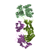
| Structure viewer | Molecule: MolmilJmol/JSmol |
|---|
- Downloads & links
-Download
| PDBx/mmCIF format | 4ogx.cif.gz | 147.7 KB | Display | PDBx/mmCIF format |
|---|---|---|---|---|
| PDB format | pdb4ogx.ent.gz | 113.5 KB | Display | PDB format |
| PDBx/mmJSON format | 4ogx.json.gz | Tree view | PDBx/mmJSON format | |
| Others |  Other downloads Other downloads |
-Validation report
| Arichive directory | https://data.pdbj.org/pub/pdb/validation_reports/og/4ogxftp://data.pdbj.org/pub/pdb/validation_reports/og/4ogx | HTTPS FTP |
|---|
-Related structure data
| Related structure data |  4ogyC  4pubC  2anyS S: Starting model for refinement C: citing same article ( |
|---|---|
| Similar structure data |


-Links
 PDBj
PDBj


- Assembly
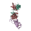
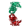
| Deposited unit | 
| ||||||||
|---|---|---|---|---|---|---|---|---|---|
| 1 |
| ||||||||
| Unit cell |
|
-Components
| #1: Protein | / Fletcher factor / Kininogenin / Plasma prekallikrein / Plasma kallikrein heavy chain / Plasma ...Fletcher factor / Kininogenin / Plasma prekallikrein / Plasma kallikrein heavy chain / Plasma kallikrein light chain Mass: 27144.842 Da / Num. of mol.: 1 / Fragment: UNP residues 391-631 / Mutation: N396E, N453E, N494E, C503S Source method: isolated from a genetically manipulated source Source: (gene. exp.) Homo sapiens (human) / Gene: KLK3, KLKB1, KLKB1_HUMAN / Cell line (production host): SF9 / Production host:   Spodoptera frugiperda (fall armyworm) / References: UniProt: P03952, plasma kallikrein Spodoptera frugiperda (fall armyworm) / References: UniProt: P03952, plasma kallikrein | ||
|---|---|---|---|
| #2: Antibody | Mass: 24039.129 Da / Num. of mol.: 1 Source method: isolated from a genetically manipulated source Source: (gene. exp.) Homo sapiens (human) / Production host:  Escherichia coli (E. coli) / References: UniProt: S6C4R2*PLUS Escherichia coli (E. coli) / References: UniProt: S6C4R2*PLUS | ||
| #3: Antibody | Mass: 23446.020 Da / Num. of mol.: 1 Source method: isolated from a genetically manipulated source Source: (gene. exp.) Homo sapiens (human) / Production host: Escherichia coli (E. coli) / References: UniProt: Q7Z3Y4*PLUS | ||
| #4: Chemical | ChemComp-SO4 / Sulfate  Mass: 96.063 Da / Num. of mol.: 6 / Source method: obtained synthetically / Formula: SO4 Mass: 96.063 Da / Num. of mol.: 6 / Source method: obtained synthetically / Formula: SO4#5: Water | ChemComp-HOH / | Water Mass: 18.015 Da / Num. of mol.: 270 / Source method: isolated from a natural source / Formula: H2O Mass: 18.015 Da / Num. of mol.: 270 / Source method: isolated from a natural source / Formula: H2O |
-Experimental details
-Experiment
| Experiment | Method: X-RAY DIFFRACTION / Number of used crystals: 1 |
|---|
- Sample preparation
| Crystal | Density Matthews: 2.67 Å3/Da / Density % sol: 54 % |
|---|---|
| Crystal grow | Temperature: 289 K / Method: vapor diffusion, sitting drop / pH: 6.5 Details: FAB DX-2930 AND HUMAN PLASMA KALLIKREIN VCID 7481 AT 150 UM OR 11.7 MG/ML AGAINST WIZ3-4 screen condition H7, 30% MPD, 10% PEG 3350, 0.1 M imidazole pH 6.5, 0.2 M ammonium sulfate, unique ...Details: FAB DX-2930 AND HUMAN PLASMA KALLIKREIN VCID 7481 AT 150 UM OR 11.7 MG/ML AGAINST WIZ3-4 screen condition H7, 30% MPD, 10% PEG 3350, 0.1 M imidazole pH 6.5, 0.2 M ammonium sulfate, unique puck ID QJO1-12, crystal tracking ID 242118H7 , VAPOR DIFFUSION, SITTING DROP, temperature 289K |
-Data collection
| Diffraction | Mean temperature: 100 K |
|---|---|
| Diffraction source | Source: ROTATING ANODE / Type: RIGAKU FR-E+ SUPERBRIGHT / Wavelength: 1.5418 Å |
| Detector | Type: RIGAKU SATURN 944+ / Detector: CCD / Date: Feb 21, 2013 / Details: VARIMAX |
| Radiation | Protocol: SINGLE WAVELENGTH / Monochromatic (M) / Laue (L): M / Scattering type: x-ray |
| Radiation wavelength | Wavelength: 1.5418 Å / Relative weight: 1 |
| Reflection | Resolution: 2.4→50 Å / Num. all: 32254 / Num. obs: 32218 / % possible obs: 99.9 % / Observed criterion σ(I): -3 / Redundancy: 6.7 % / Biso Wilson estimate: 30.87 Å2 / Rmerge(I) obs: 0.112 / Net I/σ(I): 15.74 |
| Reflection shell | Resolution: 2.4→2.45 Å / Redundancy: 4 % / Rmerge(I) obs: 0.477 / Mean I/σ(I) obs: 2.9 / Num. measured obs: 2422 / Num. unique obs: 426 / % possible all: 99.6 |
-Phasing
| Phasing | Method: molecular replacement |
|---|
- Processing
| Software |
| ||||||||||||||||||||||||||||||||||||||||||||||||||||||||||||||||||||||||||||||||||||||||||||||||||||||||||||||||||||||||||||||||||||||||||||||||||||||||||||||||||||||||||
|---|---|---|---|---|---|---|---|---|---|---|---|---|---|---|---|---|---|---|---|---|---|---|---|---|---|---|---|---|---|---|---|---|---|---|---|---|---|---|---|---|---|---|---|---|---|---|---|---|---|---|---|---|---|---|---|---|---|---|---|---|---|---|---|---|---|---|---|---|---|---|---|---|---|---|---|---|---|---|---|---|---|---|---|---|---|---|---|---|---|---|---|---|---|---|---|---|---|---|---|---|---|---|---|---|---|---|---|---|---|---|---|---|---|---|---|---|---|---|---|---|---|---|---|---|---|---|---|---|---|---|---|---|---|---|---|---|---|---|---|---|---|---|---|---|---|---|---|---|---|---|---|---|---|---|---|---|---|---|---|---|---|---|---|---|---|---|---|---|---|---|---|
| Refinement | Method to determine structure: MOLECULAR REPLACEMENT Starting model: PDB ENTRY 2ANY Resolution: 2.4→41.12 Å / Cor.coef. Fo:Fc: 0.938 / Cor.coef. Fo:Fc free: 0.901 / SU B: 6.883 / SU ML: 0.162 / Cross valid method: THROUGHOUT / σ(F): 0 / ESU R: 0.344 / ESU R Free: 0.241 / Stereochemistry target values: MAXIMUM LIKELIHOOD Details: HYDROGENS HAVE BEEN ADDED IN THE RIDING POSITIONS U VALUES : REFINED INDIVIDUALLY
| ||||||||||||||||||||||||||||||||||||||||||||||||||||||||||||||||||||||||||||||||||||||||||||||||||||||||||||||||||||||||||||||||||||||||||||||||||||||||||||||||||||||||||
| Solvent computation | Ion probe radii: 0.8 Å / Shrinkage radii: 0.8 Å / VDW probe radii: 1.2 Å / Solvent model: MASK | ||||||||||||||||||||||||||||||||||||||||||||||||||||||||||||||||||||||||||||||||||||||||||||||||||||||||||||||||||||||||||||||||||||||||||||||||||||||||||||||||||||||||||
| Displacement parameters | Biso max: 78.45 Å2 / Biso mean: 25.07 Å2 / Biso min: 3.9 Å2
| ||||||||||||||||||||||||||||||||||||||||||||||||||||||||||||||||||||||||||||||||||||||||||||||||||||||||||||||||||||||||||||||||||||||||||||||||||||||||||||||||||||||||||
| Refinement step | Cycle: LAST / Resolution: 2.4→41.12 Å
| ||||||||||||||||||||||||||||||||||||||||||||||||||||||||||||||||||||||||||||||||||||||||||||||||||||||||||||||||||||||||||||||||||||||||||||||||||||||||||||||||||||||||||
| Refine LS restraints |
| ||||||||||||||||||||||||||||||||||||||||||||||||||||||||||||||||||||||||||||||||||||||||||||||||||||||||||||||||||||||||||||||||||||||||||||||||||||||||||||||||||||||||||
| LS refinement shell | Resolution: 2.4→2.46 Å / Total num. of bins used: 20
|
