 Movie
Movie Controller

[English] 日本語
 Yorodumi
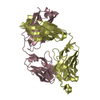
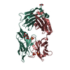
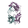
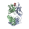
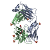
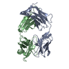
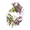
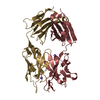
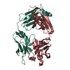
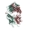
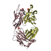
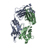
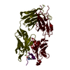
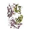
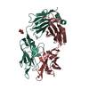
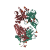
- PDB-4o4y: Crystal structure of the anti-hinge rabbit antibody 2095-2 in com... -
+ Open data

- Basic information
| Entry | Database: PDB / ID: 4o4y | |||||||||
|---|---|---|---|---|---|---|---|---|---|---|
| Title | Crystal structure of the anti-hinge rabbit antibody 2095-2 in complex with IDES hinge peptide | |||||||||
 Components Components |
| |||||||||
 Keywords Keywords |  IMMUNE SYSTEM / immunoglobulin fold / antibody / hinge IMMUNE SYSTEM / immunoglobulin fold / antibody / hinge | |||||||||
| Function / homology |  Function and homology informationcomplement-dependent cytotoxicity / antibody-dependent cellular cytotoxicity / Fc-gamma receptor I complex binding / Classical antibody-mediated complement activation / Initial triggering of complement / immunoglobulin complex, circulating / IgG immunoglobulin complex / immunoglobulin receptor binding / FCGR activation / Role of phospholipids in phagocytosis ...complement-dependent cytotoxicity / antibody-dependent cellular cytotoxicity / Fc-gamma receptor I complex binding / Classical antibody-mediated complement activation / Initial triggering of complement / immunoglobulin complex, circulating / IgG immunoglobulin complex / immunoglobulin receptor binding / FCGR activation / Role of phospholipids in phagocytosis / complement activation, classical pathway / antigen binding / FCGR3A-mediated IL10 synthesis / Regulation of Complement cascade / FCGR3A-mediated phagocytosis / B cell receptor signaling pathway / Regulation of actin dynamics for phagocytic cup formation / antibacterial humoral response / Interleukin-4 and Interleukin-13 signaling / blood microparticle / adaptive immune response / extracellular space / extracellular exosome / extracellular region / plasma membrane Function and homology informationcomplement-dependent cytotoxicity / antibody-dependent cellular cytotoxicity / Fc-gamma receptor I complex binding / Classical antibody-mediated complement activation / Initial triggering of complement / immunoglobulin complex, circulating / IgG immunoglobulin complex / immunoglobulin receptor binding / FCGR activation / Role of phospholipids in phagocytosis ...complement-dependent cytotoxicity / antibody-dependent cellular cytotoxicity / Fc-gamma receptor I complex binding / Classical antibody-mediated complement activation / Initial triggering of complement / immunoglobulin complex, circulating / IgG immunoglobulin complex / immunoglobulin receptor binding / FCGR activation / Role of phospholipids in phagocytosis / complement activation, classical pathway / antigen binding / FCGR3A-mediated IL10 synthesis / Regulation of Complement cascade / FCGR3A-mediated phagocytosis / B cell receptor signaling pathway / Regulation of actin dynamics for phagocytic cup formation / antibacterial humoral response / Interleukin-4 and Interleukin-13 signaling / blood microparticle / adaptive immune response / extracellular space / extracellular exosome / extracellular region / plasma membraneSimilarity search - Function | |||||||||
| Biological species |  Oryctolagus cuniculus (rabbit) Oryctolagus cuniculus (rabbit) Homo sapiens (human) Homo sapiens (human) | |||||||||
| Method | X-RAY DIFFRACTION / MOLECULAR REPLACEMENT / Resolution: 1.85 Å | |||||||||
 Authors Authors | Malia, T.J. / Teplyakov, A. / Luo, J. / Gilliland, G.L. | |||||||||
 Citation Citation | Journal: Proteins / Year: 2014 Title: Structure and specificity of an antibody targeting a proteolytically cleaved IgG hinge. Authors: Malia, T.J. / Teplyakov, A. / Brezski, R.J. / Luo, J. / Kinder, M. / Sweet, R.W. / Almagro, J.C. / Jordan, R.E. / Gilliland, G.L. | |||||||||
| History |
|
- Structure visualization
| Structure viewer | Molecule: MolmilJmol/JSmol |
|---|
- Downloads & links
-Download
| PDBx/mmCIF format | 4o4y.cif.gz | 197.2 KB | Display | PDBx/mmCIF format |
|---|---|---|---|---|
| PDB format | pdb4o4y.ent.gz | 156.3 KB | Display | PDB format |
| PDBx/mmJSON format | 4o4y.json.gz | Tree view | PDBx/mmJSON format | |
| Others |  Other downloads Other downloads |
-Validation report
| Arichive directory | https://data.pdbj.org/pub/pdb/validation_reports/o4/4o4yftp://data.pdbj.org/pub/pdb/validation_reports/o4/4o4y | HTTPS FTP |
|---|
-Related structure data
| Related structure data |  4ma3SC  4o51C S: Starting model for refinement C: citing same article ( |
|---|---|
| Similar structure data |


-Links
 PDBj
PDBj


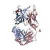
- Assembly
| Deposited unit | 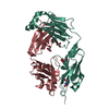
| |||||||||
|---|---|---|---|---|---|---|---|---|---|---|
| 1 |
| |||||||||
| Unit cell |
| |||||||||
| Components on special symmetry positions |
|
-Components
| #1: Antibody | Mass: 23724.293 Da / Num. of mol.: 1 / Fragment: SEE REMARK 999 Source method: isolated from a genetically manipulated source Source: (gene. exp.) Oryctolagus cuniculus, Homo sapiens / Production host: Homo sapiens (human) | ||||
|---|---|---|---|---|---|
| #2: Antibody | Mass: 23565.375 Da / Num. of mol.: 1 Source method: isolated from a genetically manipulated source Source: (gene. exp.) Oryctolagus cuniculus, Homo sapiens / Production host: Homo sapiens (human) | ||||
| #3: Protein/peptide | Mass: 1404.543 Da / Num. of mol.: 1 / Source method: obtained synthetically / Source: (synth.) Homo sapiens (human) / References: UniProt: P01857*PLUS | ||||
| #4: Chemical | ChemComp-GOL / Glycerol  Mass: 92.094 Da / Num. of mol.: 5 / Source method: obtained synthetically / Formula: C3H8O3 Mass: 92.094 Da / Num. of mol.: 5 / Source method: obtained synthetically / Formula: C3H8O3#5: Water | ChemComp-HOH / | Water Mass: 18.015 Da / Num. of mol.: 492 / Source method: isolated from a natural source / Formula: H2O Mass: 18.015 Da / Num. of mol.: 492 / Source method: isolated from a natural source / Formula: H2OSequence details | RESIDUE L CYS 300 IS A CYSTEINYLA | |
-Experimental details
-Experiment
| Experiment | Method: X-RAY DIFFRACTION / Number of used crystals: 1 |
|---|
- Sample preparation
| Crystal | Density Matthews: 2.46 Å3/Da / Density % sol: 50.09 % |
|---|---|
| Crystal grow | Temperature: 293 K / Method: vapor diffusion, sitting drop / pH: 8.5 Details: 0.1 M Tris, pH 8.5, 18.33% PEG3350, 0.2 M lithium sulfate, 5% isopropanol, cryoprotection: mother liquor + 20% glycerol, VAPOR DIFFUSION, SITTING DROP, temperature 293.0K |
-Data collection
| Diffraction | Mean temperature: 100 K |
|---|---|
| Diffraction source | Source: ROTATING ANODE / Type: RIGAKU MICROMAX-007 HF / Wavelength: 1.5418 / Wavelength: 1.5418 Å |
| Detector | Type: RIGAKU SATURN 944 / Detector: CCD / Date: Dec 16, 2009 / Details: VARIMAX HF |
| Radiation | Protocol: SINGLE WAVELENGTH / Monochromatic (M) / Laue (L): M / Scattering type: x-ray |
| Radiation wavelength | Wavelength: 1.5418 Å / Relative weight: 1 |
| Reflection | Resolution: 1.85→25 Å / Num. obs: 37290 / % possible obs: 89.2 % / Observed criterion σ(F): 0 / Observed criterion σ(I): -3 / Redundancy: 8.9 % / Biso Wilson estimate: 29 Å2 / Rmerge(I) obs: 0.059 / Net I/σ(I): 17.5 |
| Reflection shell | Resolution: 1.85→1.92 Å / Redundancy: 6.9 % / Rmerge(I) obs: 0.242 / Mean I/σ(I) obs: 6.1 / % possible all: 51.6 |
- Processing
| Software |
| ||||||||||||||||||||||||||||||||||||||||||||||||||||||||||||||||||||||||||||||||||||||||||||||||||||||||||||||||||||||||||||||||||||||||||||||||||||||
|---|---|---|---|---|---|---|---|---|---|---|---|---|---|---|---|---|---|---|---|---|---|---|---|---|---|---|---|---|---|---|---|---|---|---|---|---|---|---|---|---|---|---|---|---|---|---|---|---|---|---|---|---|---|---|---|---|---|---|---|---|---|---|---|---|---|---|---|---|---|---|---|---|---|---|---|---|---|---|---|---|---|---|---|---|---|---|---|---|---|---|---|---|---|---|---|---|---|---|---|---|---|---|---|---|---|---|---|---|---|---|---|---|---|---|---|---|---|---|---|---|---|---|---|---|---|---|---|---|---|---|---|---|---|---|---|---|---|---|---|---|---|---|---|---|---|---|---|---|---|---|---|
| Refinement | Method to determine structure: MOLECULAR REPLACEMENT Starting model: PDB ENTRY 4MA3 Resolution: 1.85→25 Å / Cor.coef. Fo:Fc: 0.956 / Cor.coef. Fo:Fc free: 0.937 / Occupancy max: 1 / Occupancy min: 0.21 / SU B: 6.217 / SU ML: 0.086 / Cross valid method: THROUGHOUT / σ(F): 0 / ESU R: 0.169 / ESU R Free: 0.155 / Stereochemistry target values: MAXIMUM LIKELIHOOD / Details: HYDROGENS HAVE BEEN ADDED IN THE RIDING POSITIONS
| ||||||||||||||||||||||||||||||||||||||||||||||||||||||||||||||||||||||||||||||||||||||||||||||||||||||||||||||||||||||||||||||||||||||||||||||||||||||
| Solvent computation | Ion probe radii: 0.8 Å / Shrinkage radii: 0.8 Å / VDW probe radii: 1.4 Å / Solvent model: MASK | ||||||||||||||||||||||||||||||||||||||||||||||||||||||||||||||||||||||||||||||||||||||||||||||||||||||||||||||||||||||||||||||||||||||||||||||||||||||
| Displacement parameters | Biso max: 117.79 Å2 / Biso mean: 33.146 Å2 / Biso min: 5.39 Å2
| ||||||||||||||||||||||||||||||||||||||||||||||||||||||||||||||||||||||||||||||||||||||||||||||||||||||||||||||||||||||||||||||||||||||||||||||||||||||
| Refinement step | Cycle: LAST / Resolution: 1.85→25 Å
| ||||||||||||||||||||||||||||||||||||||||||||||||||||||||||||||||||||||||||||||||||||||||||||||||||||||||||||||||||||||||||||||||||||||||||||||||||||||
| Refine LS restraints |
| ||||||||||||||||||||||||||||||||||||||||||||||||||||||||||||||||||||||||||||||||||||||||||||||||||||||||||||||||||||||||||||||||||||||||||||||||||||||
| LS refinement shell | Resolution: 1.85→1.898 Å / Total num. of bins used: 20
| ||||||||||||||||||||||||||||||||||||||||||||||||||||||||||||||||||||||||||||||||||||||||||||||||||||||||||||||||||||||||||||||||||||||||||||||||||||||
| Refinement TLS params. | Method: refined / Refine-ID: X-RAY DIFFRACTION
| ||||||||||||||||||||||||||||||||||||||||||||||||||||||||||||||||||||||||||||||||||||||||||||||||||||||||||||||||||||||||||||||||||||||||||||||||||||||
| Refinement TLS group |
|
