 Movie
Movie Controller


+ Open data

- Basic information
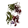
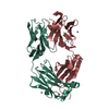
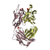
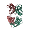
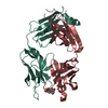
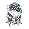
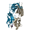
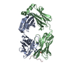
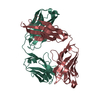
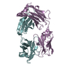
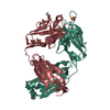
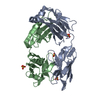
| Entry | Database: PDB / ID: 3i75 | ||||||
|---|---|---|---|---|---|---|---|
| Title | Antibody Structure | ||||||
 Components Components |
| ||||||
 Keywords Keywords |  IMMUNE SYSTEM / Antibody / IgG IMMUNE SYSTEM / Antibody / IgG | ||||||
| Function / homology | Immunoglobulins / Immunoglobulin-like / Sandwich / Mainly Beta Function and homology information Function and homology information | ||||||
| Biological species |  Mus musculus (house mouse) Mus musculus (house mouse) | ||||||
| Method | X-RAY DIFFRACTION / SYNCHROTRON / MOLECULAR REPLACEMENT / Resolution: 1.95 Å | ||||||
 Authors Authors | Isaacs, N.W. / Riboldi-Tunnicliffe, A. | ||||||
 Citation Citation | Journal: To be Published Title: Antibody Structure Authors: Riboldi-Tunnicliffe, A. / Isaacs, N.W. | ||||||
| History |
|
- Structure visualization
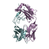
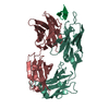
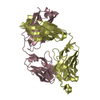
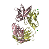
| Structure viewer | Molecule: MolmilJmol/JSmol |
|---|
- Downloads & links
-Download
| PDBx/mmCIF format | 3i75.cif.gz | 187.3 KB | Display | PDBx/mmCIF format |
|---|---|---|---|---|
| PDB format | pdb3i75.ent.gz | 149.4 KB | Display | PDB format |
| PDBx/mmJSON format | 3i75.json.gz | Tree view | PDBx/mmJSON format | |
| Others |  Other downloads Other downloads |
-Validation report
| Arichive directory | https://data.pdbj.org/pub/pdb/validation_reports/i7/3i75ftp://data.pdbj.org/pub/pdb/validation_reports/i7/3i75 | HTTPS FTP |
|---|
-Related structure data
| Related structure data |  1mimS S: Starting model for refinement |
|---|---|
| Similar structure data |


-Links
 PDBj
PDBj

- Assembly
| Deposited unit | 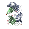
| |||||||||
|---|---|---|---|---|---|---|---|---|---|---|
| 1 |
| |||||||||
| Unit cell |
| |||||||||
| Components on special symmetry positions |
|
-Components
-Antibody , 2 types, 2 molecules AB
| #1: Antibody | Immunoglobulin heavy chain Mass: 23449.949 Da / Num. of mol.: 1 / Source method: isolated from a natural source / Source: (natural) Mus musculus (house mouse) |
|---|---|
| #2: Antibody | Immunoglobulin light chain Mass: 24125.871 Da / Num. of mol.: 1 / Source method: isolated from a natural source / Source: (natural) Mus musculus (house mouse) |
-Non-polymers , 4 types, 261 molecules 


| #3: Chemical | ChemComp-SO4 / Sulfate Mass: 96.063 Da / Num. of mol.: 6 / Source method: obtained synthetically / Formula: SO4 Mass: 96.063 Da / Num. of mol.: 6 / Source method: obtained synthetically / Formula: SO4#4: Chemical | ChemComp-CL / | Chloride Mass: 35.453 Da / Num. of mol.: 1 / Source method: obtained synthetically / Formula: Cl Mass: 35.453 Da / Num. of mol.: 1 / Source method: obtained synthetically / Formula: Cl#5: Chemical | Glycerol Mass: 92.094 Da / Num. of mol.: 2 / Source method: obtained synthetically / Formula: C3H8O3 Mass: 92.094 Da / Num. of mol.: 2 / Source method: obtained synthetically / Formula: C3H8O3#6: Water | ChemComp-HOH / | WaterMass: 18.015 Da / Num. of mol.: 252 / Source method: isolated from a natural source / Formula: H2O |
|---|
-Details
| Sequence details | AUTHOR DID NOT PROVIDE EXACT SEQUENCE FOR RESIDUES 140 AND 141 IN CHAIN B. |
|---|
-Experimental details
-Experiment
| Experiment | Method: X-RAY DIFFRACTION |
|---|
- Sample preparation
| Crystal | Density Matthews: 3.8 Å3/Da / Density % sol: 67.63 % |
|---|---|
| Crystal grow | Method: vapor diffusion / Details: VAPOR DIFFUSION |
-Data collection
| Diffraction source | Source: SYNCHROTRON / Site: SRS  / Beamline: PX7.2 / Beamline: PX7.2 |
|---|---|
| Detector | Type: MAC Science DIP-2030 / Detector: IMAGE PLATE / Date: Jun 1, 1998 |
| Radiation | Protocol: SINGLE WAVELENGTH / Scattering type: x-ray |
| Radiation wavelength | Relative weight: 1 |
| Reflection | Resolution: 1.95→111 Å / Num. obs: 51886 |
- Processing
| Software | Name: REFMAC / Version: 5.1.24 / Classification: refinement | |||||||||||||||||||||||||||||||||||||||||||||||||||||||||||||||||||||||||||||||||||||||||||||||||||||||||||||||||||
|---|---|---|---|---|---|---|---|---|---|---|---|---|---|---|---|---|---|---|---|---|---|---|---|---|---|---|---|---|---|---|---|---|---|---|---|---|---|---|---|---|---|---|---|---|---|---|---|---|---|---|---|---|---|---|---|---|---|---|---|---|---|---|---|---|---|---|---|---|---|---|---|---|---|---|---|---|---|---|---|---|---|---|---|---|---|---|---|---|---|---|---|---|---|---|---|---|---|---|---|---|---|---|---|---|---|---|---|---|---|---|---|---|---|---|---|---|
| Refinement | Method to determine structure: MOLECULAR REPLACEMENT Starting model: PDB ENTRY 1MIM Resolution: 1.95→64.83 Å / Cor.coef. Fo:Fc: 0.966 / Cor.coef. Fo:Fc free: 0.956 / SU B: 3.791 / SU ML: 0.099 / Cross valid method: THROUGHOUT / ESU R: 0.177 / ESU R Free: 0.118 / Stereochemistry target values: MAXIMUM LIKELIHOOD / Details: HYDROGENS HAVE BEEN ADDED IN THE RIDING POSITIONS
| |||||||||||||||||||||||||||||||||||||||||||||||||||||||||||||||||||||||||||||||||||||||||||||||||||||||||||||||||||
| Solvent computation | Ion probe radii: 0.8 Å / Shrinkage radii: 0.8 Å / VDW probe radii: 1.4 Å / Solvent model: BABINET MODEL WITH MASK | |||||||||||||||||||||||||||||||||||||||||||||||||||||||||||||||||||||||||||||||||||||||||||||||||||||||||||||||||||
| Displacement parameters | Biso mean: 33.778 Å2
| |||||||||||||||||||||||||||||||||||||||||||||||||||||||||||||||||||||||||||||||||||||||||||||||||||||||||||||||||||
| Refinement step | Cycle: LAST / Resolution: 1.95→64.83 Å
| |||||||||||||||||||||||||||||||||||||||||||||||||||||||||||||||||||||||||||||||||||||||||||||||||||||||||||||||||||
| Refine LS restraints |
| |||||||||||||||||||||||||||||||||||||||||||||||||||||||||||||||||||||||||||||||||||||||||||||||||||||||||||||||||||
| LS refinement shell | Resolution: 1.954→2.005 Å / Total num. of bins used: 20 /
|
