 Movie
Movie Controller


+ Open data

- Basic information
| Entry | Database: PDB / ID: 4nsq | ||||||
|---|---|---|---|---|---|---|---|
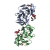
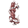
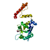
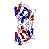
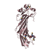
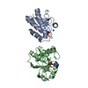
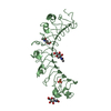
| Title | Crystal structure of PCAF | ||||||
 Components Components | Histone acetyltransferase KAT2B | ||||||
 Keywords Keywords |  TRANSFERASE / acetyltransferase / COA Binding TRANSFERASE / acetyltransferase / COA Binding | ||||||
| Function / homology |  Function and homology information Function and homology informationregulation of protein ADP-ribosylation / negative regulation of rRNA processing / histone H3K9 acetyltransferase activity / diamine N-acetyltransferase / diamine N-acetyltransferase activity / positive regulation of transcription from RNA polymerase II promoter by glucose / negative regulation of centriole replication / Physiological factors / positive regulation of attachment of mitotic spindle microtubules to kinetochore / peptidyl-lysine acetylation ...regulation of protein ADP-ribosylation / negative regulation of rRNA processing / histone H3K9 acetyltransferase activity / diamine N-acetyltransferase / diamine N-acetyltransferase activity / positive regulation of transcription from RNA polymerase II promoter by glucose / negative regulation of centriole replication / Physiological factors / positive regulation of attachment of mitotic spindle microtubules to kinetochore / peptidyl-lysine acetylation / lysine N-acetyltransferase activity, acting on acetyl phosphate as donor / YAP1- and WWTR1 (TAZ)-stimulated gene expression / histone H3 acetyltransferase activity / positive regulation of fatty acid biosynthetic process / actomyosin / internal peptidyl-lysine acetylation / cyclin-dependent protein serine/threonine kinase inhibitor activity / ATAC complex / N-terminal peptidyl-lysine acetylation / I band / cellular response to parathyroid hormone stimulus / Regulation of gene expression in late stage (branching morphogenesis) pancreatic bud precursor cells / SAGA complex / RUNX3 regulates NOTCH signaling / limb development / A band / NOTCH4 Intracellular Domain Regulates Transcription / Regulation of FOXO transcriptional activity by acetylation / NOTCH3 Intracellular Domain Regulates Transcription / regulation of tubulin deacetylation / peptide-lysine-N-acetyltransferase activity / histone acetyltransferase binding / protein acetylation / Notch-HLH transcription pathway / Formation of WDR5-containing histone-modifying complexes / regulation of cell division / Formation of paraxial mesoderm / regulation of RNA splicing / acetyltransferase activity / RNA Polymerase I Transcription Initiation / regulation of embryonic development / regulation of DNA repair / histone acetyltransferase activity / positive regulation of gluconeogenesis / histone acetyltransferase / transcription initiation-coupled chromatin remodeling / positive regulation of glycolytic process / gluconeogenesis / transcription coregulator activity / RNA polymerase II transcription regulatory region sequence-specific DNA binding / RUNX1 regulates genes involved in megakaryocyte differentiation and platelet function / B-WICH complex positively regulates rRNA expression / NOTCH1 Intracellular Domain Regulates Transcription / Metalloprotease DUBs / kinetochore / mitotic spindle / memory / Pre-NOTCH Transcription and Translation / Constitutive Signaling by NOTCH1 PEST Domain Mutants / Constitutive Signaling by NOTCH1 HD+PEST Domain Mutants / positive regulation of neuron projection development / cellular response to insulin stimulus / vasodilation / histone deacetylase binding / rhythmic process / cellular response to oxidative stress / heart development / HATs acetylate histones / DNA-binding transcription factor binding / Estrogen-dependent gene expression / transcription coactivator activity / regulation of cell cycle / chromatin remodeling / cell cycle / negative regulation of cell population proliferation / centrosome / chromatin binding / regulation of DNA-templated transcription / regulation of transcription by RNA polymerase II / protein kinase binding / positive regulation of DNA-templated transcription / negative regulation of transcription by RNA polymerase II / positive regulation of transcription by RNA polymerase II / protein-containing complex / nucleoplasm / nucleus / cytosolSimilarity search - Function | ||||||
| Biological species |  Homo sapiens (human) Homo sapiens (human) | ||||||
| Method | X-RAY DIFFRACTION / SYNCHROTRON / MOLECULAR REPLACEMENT / Resolution: 2.3108 Å | ||||||
 Authors Authors | Lin, J.Y. / Cai, Y.F. | ||||||
 Citation Citation | Journal: Bmc Struct.Biol. / Year: 2014 Title: Dimeric structure of p300/CBP associated factor. Authors: Shi, S. / Lin, J. / Cai, Y. / Yu, J. / Hong, H. / Ji, K. / Downey, J.S. / Lu, X. / Chen, R. / Han, J. / Han, A. | ||||||
| History |
|
- Structure visualization
| Structure viewer | Molecule: MolmilJmol/JSmol |
|---|
- Downloads & links
-Download
| PDBx/mmCIF format | 4nsq.cif.gz | 141.6 KB | Display | PDBx/mmCIF format |
|---|---|---|---|---|
| PDB format | pdb4nsq.ent.gz | 112.7 KB | Display | PDB format |
| PDBx/mmJSON format | 4nsq.json.gz | Tree view | PDBx/mmJSON format | |
| Others |  Other downloads Other downloads |
-Validation report
| Arichive directory | https://data.pdbj.org/pub/pdb/validation_reports/ns/4nsqftp://data.pdbj.org/pub/pdb/validation_reports/ns/4nsq | HTTPS FTP |
|---|
-Related structure data
| Similar structure data |
|---|


-Links
 PDBj
PDBj


- Assembly
| Deposited unit | 
| ||||||||
|---|---|---|---|---|---|---|---|---|---|
| 1 | 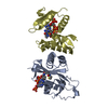
| ||||||||
| 2 | 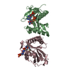
| ||||||||
| Unit cell |
|
-Components
| #1: Protein | Mass: 21838.416 Da / Num. of mol.: 4 / Fragment: UNP Residues 493-658 Source method: isolated from a genetically manipulated source Source: (gene. exp.) Homo sapiens (human) / Gene: KAT2B, PCAF / Production host:  Escherichia coli (E. coli) / References: UniProt: Q92831, histone acetyltransferase Escherichia coli (E. coli) / References: UniProt: Q92831, histone acetyltransferase#2: Chemical | ChemComp-COA / Coenzyme A  Mass: 767.534 Da / Num. of mol.: 4 / Source method: obtained synthetically / Formula: C21H36N7O16P3S Mass: 767.534 Da / Num. of mol.: 4 / Source method: obtained synthetically / Formula: C21H36N7O16P3S |
|---|
-Experimental details
-Experiment
| Experiment | Method: X-RAY DIFFRACTION / Number of used crystals: 1 |
|---|
- Sample preparation
| Crystal | Density Matthews: 2.31 Å3/Da / Density % sol: 46.72 % |
|---|---|
| Crystal grow | Temperature: 298 K / Method: vapor diffusion / pH: 7.4 Details: 0.2M Lithium Sulfate, 0.1M HEPES pH7.4, 1.2M Ammonium Sulfate and 10mM Trimethylamine HCl, VAPOR DIFFUSION, temperature 298K |
-Data collection
| Diffraction | Mean temperature: 100 K | ||||||||||||||||||
|---|---|---|---|---|---|---|---|---|---|---|---|---|---|---|---|---|---|---|---|
| Diffraction source | Source: SYNCHROTRON / Site: SSRF  / Beamline: BL17U / Wavelength: 0.97916 Å / Beamline: BL17U / Wavelength: 0.97916 Å | ||||||||||||||||||
| Detector | Type: MARMOSAIC 225 mm CCD / Detector: CCD / Date: May 24, 2010 | ||||||||||||||||||
| Radiation | Monochromator: Si 111 / Protocol: SINGLE WAVELENGTH / Monochromatic (M) / Laue (L): M / Scattering type: x-ray | ||||||||||||||||||
| Radiation wavelength | Wavelength: 0.97916 Å / Relative weight: 1 | ||||||||||||||||||
| Reflection | Resolution: 2.31→50 Å / Num. obs: 30459 / % possible obs: 99.3 % / Observed criterion σ(F): 3 / Observed criterion σ(I): 3 | ||||||||||||||||||
| Reflection shell |
|
- Processing
| Software |
| ||||||||||||||||||||||||||||||||||||||||||||||||||||||||||||||||||||||||||||||||||||||||||||||||||
|---|---|---|---|---|---|---|---|---|---|---|---|---|---|---|---|---|---|---|---|---|---|---|---|---|---|---|---|---|---|---|---|---|---|---|---|---|---|---|---|---|---|---|---|---|---|---|---|---|---|---|---|---|---|---|---|---|---|---|---|---|---|---|---|---|---|---|---|---|---|---|---|---|---|---|---|---|---|---|---|---|---|---|---|---|---|---|---|---|---|---|---|---|---|---|---|---|---|---|---|
| Refinement | Method to determine structure: MOLECULAR REPLACEMENT / Resolution: 2.3108→45.256 Å / SU ML: 0.73 / σ(F): 2 / Phase error: 35.03 / Stereochemistry target values: ML
| ||||||||||||||||||||||||||||||||||||||||||||||||||||||||||||||||||||||||||||||||||||||||||||||||||
| Solvent computation | Shrinkage radii: 0.83 Å / VDW probe radii: 1.1 Å / Solvent model: FLAT BULK SOLVENT MODEL / Bsol: 49.081 Å2 / ksol: 0.324 e/Å3 | ||||||||||||||||||||||||||||||||||||||||||||||||||||||||||||||||||||||||||||||||||||||||||||||||||
| Displacement parameters |
| ||||||||||||||||||||||||||||||||||||||||||||||||||||||||||||||||||||||||||||||||||||||||||||||||||
| Refinement step | Cycle: LAST / Resolution: 2.3108→45.256 Å
| ||||||||||||||||||||||||||||||||||||||||||||||||||||||||||||||||||||||||||||||||||||||||||||||||||
| Refine LS restraints |
| ||||||||||||||||||||||||||||||||||||||||||||||||||||||||||||||||||||||||||||||||||||||||||||||||||
| LS refinement shell |
|
