 Movie
Movie Controller

[English] 日本語
 Yorodumi
- PDB-4nom: Crystal structure of asparaginyl endopeptidase (AEP)/Legumain act... -
+ Open data

- Basic information
| Entry | Database: PDB / ID: 4nom | ||||||
|---|---|---|---|---|---|---|---|
| Title | Crystal structure of asparaginyl endopeptidase (AEP)/Legumain activated at pH 4.5 | ||||||
 Components Components | Legumain Asparagine endopeptidase Asparagine endopeptidase | ||||||
 Keywords Keywords | HYDROLASE / new fold / asparaginyl endopeptidase / substrate / extracellular | ||||||
| Function / homology |  Function and homology information Function and homology informationTrafficking and processing of endosomal TLR / negative regulation of ERBB signaling pathway / legumain / vacuolar protein processing / renal system process / receptor catabolic process / self proteolysis / MHC class II antigen presentation / positive regulation of endothelial cell chemotaxis / response to acidic pH ...Trafficking and processing of endosomal TLR / negative regulation of ERBB signaling pathway / legumain / vacuolar protein processing / renal system process / receptor catabolic process / self proteolysis / MHC class II antigen presentation / positive regulation of endothelial cell chemotaxis / response to acidic pH / dendritic spine organization / positive regulation of monocyte chemotaxis / negative regulation of multicellular organism growth / cellular response to hepatocyte growth factor stimulus / associative learning / protein maturation / endopeptidase activator activity / cellular response to calcium ion / proteolysis involved in protein catabolic process / positive regulation of mitotic cell cycle / lysosomal lumen / positive regulation of long-term synaptic potentiation / memory / cellular response to amyloid-beta / antigen processing and presentation of exogenous peptide antigen via MHC class II / late endosome / apical part of cell / peptidase activity / negative regulation of neuron apoptotic process / lysosome / cysteine-type endopeptidase activity / negative regulation of gene expression / positive regulation of cell population proliferation / perinuclear region of cytoplasm / proteolysis / extracellular region / cytoplasmSimilarity search - Function | ||||||
| Biological species |  Mus musculus (house mouse) Mus musculus (house mouse) | ||||||
| Method | X-RAY DIFFRACTION / SYNCHROTRON / SAD / Resolution: 2.006 Å | ||||||
 Authors Authors | Zhao, L. / Hua, T. / Ru, H. / Ni, X. / Shaw, N. / Jiao, L. / Ding, W. / Qu, L. / Ouyang, S. / Liu, Z.J. | ||||||
 Citation Citation | Journal: Cell Res. / Year: 2014 Title: Structural analysis of asparaginyl endopeptidase reveals the activation mechanism and a reversible intermediate maturation stage. Authors: Zhao, L. / Hua, T. / Crowley, C. / Ru, H. / Ni, X. / Shaw, N. / Jiao, L. / Ding, W. / Qu, L. / Hung, L.W. / Huang, W. / Liu, L. / Ye, K. / Ouyang, S. / Cheng, G. / Liu, Z.J. | ||||||
| History |
|
- Structure visualization
| Structure viewer | Molecule: MolmilJmol/JSmol |
|---|
- Downloads & links
-Download
| PDBx/mmCIF format | 4nom.cif.gz | 171.4 KB | Display | PDBx/mmCIF format |
|---|---|---|---|---|
| PDB format | pdb4nom.ent.gz | 143.9 KB | Display | PDB format |
| PDBx/mmJSON format | 4nom.json.gz | Tree view | PDBx/mmJSON format | |
| Others |  Other downloads Other downloads |
-Validation report
| Arichive directory | https://data.pdbj.org/pub/pdb/validation_reports/no/4nomftp://data.pdbj.org/pub/pdb/validation_reports/no/4nom | HTTPS FTP |
|---|
-Related structure data


-Links
 PDBj
PDBj


- Assembly
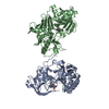
| Deposited unit | 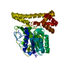
| |||||||||
|---|---|---|---|---|---|---|---|---|---|---|
| 1 | 
| |||||||||
| Unit cell |
| |||||||||
| Components on special symmetry positions |
|
-Components
| #1: Protein | Asparagine endopeptidase / Asparaginyl endopeptidase / Protease / cysteine 1 Mass: 50266.758 Da / Num. of mol.: 1 Source method: isolated from a genetically manipulated source Source: (gene. exp.) Mus musculus (house mouse) / Gene: Lgmn, Prsc1 / Production host:   Spodoptera frugiperda (fall armyworm) / References: UniProt: O89017, legumain Spodoptera frugiperda (fall armyworm) / References: UniProt: O89017, legumain |
|---|---|
| #2: Water | ChemComp-HOH / Water Mass: 18.015 Da / Num. of mol.: 158 / Source method: isolated from a natural source / Formula: H2O Mass: 18.015 Da / Num. of mol.: 158 / Source method: isolated from a natural source / Formula: H2O |
-Experimental details
-Experiment
| Experiment | Method: X-RAY DIFFRACTION / Number of used crystals: 1 |
|---|
- Sample preparation
| Crystal | Density Matthews: 2.51 Å3/Da / Density % sol: 51 % |
|---|---|
| Crystal grow | Temperature: 298 K / Method: vapor diffusion, hanging drop / pH: 8.5 Details: 0.2 M trimethylamine N-oxide dehydrate, 0.1 M Tris-HCl, pH 8.5, and 20% PEG 2,000, VAPOR DIFFUSION, HANGING DROP, temperature 298K |
-Data collection
| Diffraction | Mean temperature: 100 K |
|---|---|
| Diffraction source | Source: SYNCHROTRON / Site: SSRF  / Beamline: BL17U / Wavelength: 0.98 Å / Beamline: BL17U / Wavelength: 0.98 Å |
| Detector | Type: ADSC QUANTUM 315 / Detector: CCD / Date: Oct 1, 2012 |
| Radiation | Monochromator: GRAPHITE / Protocol: SINGLE WAVELENGTH / Monochromatic (M) / Laue (L): M / Scattering type: x-ray |
| Radiation wavelength | Wavelength: 0.98 Å / Relative weight: 1 |
| Reflection | Resolution: 2→50 Å / Num. all: 32142 / Num. obs: 32142 / % possible obs: 100 % / Observed criterion σ(F): 2 / Observed criterion σ(I): 2 |
- Processing
| Software |
| |||||||||||||||||||||||||||||||||||||||||||||||||||||||||||||||||||||||||||||||||||||||||||
|---|---|---|---|---|---|---|---|---|---|---|---|---|---|---|---|---|---|---|---|---|---|---|---|---|---|---|---|---|---|---|---|---|---|---|---|---|---|---|---|---|---|---|---|---|---|---|---|---|---|---|---|---|---|---|---|---|---|---|---|---|---|---|---|---|---|---|---|---|---|---|---|---|---|---|---|---|---|---|---|---|---|---|---|---|---|---|---|---|---|---|---|---|
| Refinement | Method to determine structure: SAD / Resolution: 2.006→32.623 Å / SU ML: 0.26 / σ(F): 1.34 / Phase error: 28.75 / Stereochemistry target values: ML
| |||||||||||||||||||||||||||||||||||||||||||||||||||||||||||||||||||||||||||||||||||||||||||
| Solvent computation | Shrinkage radii: 0.98 Å / VDW probe radii: 1.2 Å / Solvent model: FLAT BULK SOLVENT MODEL / Bsol: 37.454 Å2 / ksol: 0.329 e/Å3 | |||||||||||||||||||||||||||||||||||||||||||||||||||||||||||||||||||||||||||||||||||||||||||
| Displacement parameters |
| |||||||||||||||||||||||||||||||||||||||||||||||||||||||||||||||||||||||||||||||||||||||||||
| Refinement step | Cycle: LAST / Resolution: 2.006→32.623 Å
| |||||||||||||||||||||||||||||||||||||||||||||||||||||||||||||||||||||||||||||||||||||||||||
| Refine LS restraints |
| |||||||||||||||||||||||||||||||||||||||||||||||||||||||||||||||||||||||||||||||||||||||||||
| LS refinement shell |
| |||||||||||||||||||||||||||||||||||||||||||||||||||||||||||||||||||||||||||||||||||||||||||
| Refinement TLS params. | Method: refined / Origin x: 42.4124 Å / Origin y: 22.4794 Å / Origin z: 28.143 Å
| |||||||||||||||||||||||||||||||||||||||||||||||||||||||||||||||||||||||||||||||||||||||||||
| Refinement TLS group | Selection details: all |
