 Movie
Movie Controller

[English] 日本語
 Yorodumi
- PDB-4nf6: Crystal structure of GluN1/GluN2A ligand-binding domain in comple... -
+ Open data

- Basic information
| Entry | Database: PDB / ID: 4nf6 | ||||||
|---|---|---|---|---|---|---|---|
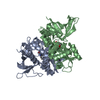
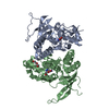
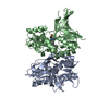
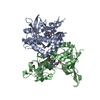
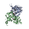
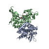
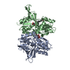
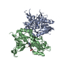
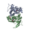
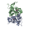
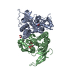
| Title | Crystal structure of GluN1/GluN2A ligand-binding domain in complex with glycine and PPDA | ||||||
 Components Components |
| ||||||
 Keywords Keywords |  transport protein / receptor / glycine and PPDA transport protein / receptor / glycine and PPDA | ||||||
| Function / homology |  Function and homology information Function and homology informationresponse to ammonium ion / directional locomotion / pons maturation / regulation of cell communication / positive regulation of Schwann cell migration / EPHB-mediated forward signaling / serotonin metabolic process / Assembly and cell surface presentation of NMDA receptors / olfactory learning / conditioned taste aversion ...response to ammonium ion / directional locomotion / pons maturation / regulation of cell communication / positive regulation of Schwann cell migration / EPHB-mediated forward signaling / serotonin metabolic process / Assembly and cell surface presentation of NMDA receptors / olfactory learning / conditioned taste aversion / protein localization to postsynaptic membrane / dendritic branch / regulation of respiratory gaseous exchange / propylene metabolic process / response to glycine / response to other organism / sleep / cellular response to magnesium ion / response to methylmercury / voltage-gated monoatomic cation channel activity / locomotion / response to morphine / glutamate-gated calcium ion channel activity / cellular response to dsRNA / response to carbohydrate / regulation of monoatomic cation transmembrane transport / dendritic spine organization / cellular response to lipid / NMDA glutamate receptor activity / NMDA selective glutamate receptor complex / RAF/MAP kinase cascade / Synaptic adhesion-like molecules / parallel fiber to Purkinje cell synapse / calcium ion transmembrane import into cytosol / response to manganese ion / protein heterotetramerization / glutamate binding / positive regulation of reactive oxygen species biosynthetic process / cellular response to zinc ion / neuromuscular process / regulation of synapse assembly / action potential / glycine binding / positive regulation of calcium ion transport into cytosol / male mating behavior / regulation of dendrite morphogenesis / regulation of axonogenesis / dopamine metabolic process / spinal cord development / suckling behavior / startle response / regulation of neuronal synaptic plasticity / response to amine / monoatomic cation transmembrane transport / regulation of NMDA receptor activity / social behavior / associative learning / positive regulation of excitatory postsynaptic potential / monoatomic cation transport / ligand-gated monoatomic ion channel activity / response to light stimulus / excitatory synapse / Unblocking of NMDA receptors, glutamate binding and activation / neuron development / positive regulation of dendritic spine maintenance / glutamate receptor binding / regulation of postsynaptic membrane potential / phosphatase binding / calcium ion homeostasis / cellular response to manganese ion / prepulse inhibition / long-term memory / regulation of neuron apoptotic process / glutamate-gated receptor activity / synaptic cleft / presynaptic active zone membrane / response to fungicide / monoatomic cation channel activity / sensory perception of pain / dendrite membrane / ligand-gated monoatomic ion channel activity involved in regulation of presynaptic membrane potential / response to amphetamine / excitatory postsynaptic potential / hippocampal mossy fiber to CA3 synapse / regulation of membrane potential / ionotropic glutamate receptor signaling pathway / cell adhesion molecule binding / neurogenesis / positive regulation of synaptic transmission, glutamatergic / adult locomotory behavior / response to cocaine / transmitter-gated monoatomic ion channel activity involved in regulation of postsynaptic membrane potential / synaptic membrane / synaptic transmission, glutamatergic / learning / long-term synaptic potentiation / hippocampus development / cellular response to amino acid stimulus / postsynaptic density membrane / regulation of long-term neuronal synaptic plasticitySimilarity search - Function | ||||||
| Biological species |  Rattus norvegicus (Norway rat) Rattus norvegicus (Norway rat) | ||||||
| Method | X-RAY DIFFRACTION / SYNCHROTRON / MOLECULAR REPLACEMENT / Resolution: 2.1 Å | ||||||
 Authors Authors | Jespersen, A. / Tajima, N. / Furukawa, H. | ||||||
 Citation Citation | Journal: Neuron / Year: 2014 Title: Structural Insights into Competitive Antagonism in NMDA Receptors. Authors: Jespersen, A. / Tajima, N. / Fernandez-Cuervo, G. / Garnier-Amblard, E.C. / Furukawa, H. | ||||||
| History |
|
- Structure visualization
| Structure viewer | Molecule: MolmilJmol/JSmol |
|---|
- Downloads & links
-Download
| PDBx/mmCIF format | 4nf6.cif.gz | 134.9 KB | Display | PDBx/mmCIF format |
|---|---|---|---|---|
| PDB format | pdb4nf6.ent.gz | 109.2 KB | Display | PDB format |
| PDBx/mmJSON format | 4nf6.json.gz | Tree view | PDBx/mmJSON format | |
| Others |  Other downloads Other downloads |
-Validation report
| Arichive directory | https://data.pdbj.org/pub/pdb/validation_reports/nf/4nf6ftp://data.pdbj.org/pub/pdb/validation_reports/nf/4nf6 | HTTPS FTP |
|---|
-Related structure data


-Links
 PDBj
PDBj


- Assembly
| Deposited unit | 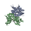
| ||||||||
|---|---|---|---|---|---|---|---|---|---|
| 1 |
| ||||||||
| Unit cell |
|
-Components
| #1: Protein | Mass: 33340.031 Da / Num. of mol.: 1 Fragment: Ligand-binding domain, unp residues 393-543; unp residues 663-800 Source method: isolated from a genetically manipulated source Source: (gene. exp.) Rattus norvegicus (Norway rat) / Gene: Grin1, Nmdar1 / Production host:  Escherichia coli (E. coli) / References: UniProt: P35439 Escherichia coli (E. coli) / References: UniProt: P35439 |
|---|---|
| #2: Protein | Mass: 31785.299 Da / Num. of mol.: 1 Fragment: Ligand-binding domain; unp residues 402-539; unp residues 661-802 Mutation: S758T Source method: isolated from a genetically manipulated source Source: (gene. exp.) Rattus norvegicus (Norway rat) / Gene: Grin2a / Production host: Escherichia coli (E. coli) / References: UniProt: Q00959 |
| #3: Chemical | ChemComp-GLY / Glycine  Type: peptide linking / Mass: 75.067 Da / Num. of mol.: 1 / Source method: obtained synthetically / Formula: C2H5NO2 Type: peptide linking / Mass: 75.067 Da / Num. of mol.: 1 / Source method: obtained synthetically / Formula: C2H5NO2 |
| #4: Chemical | ChemComp-2JL / (  Mass: 378.378 Da / Num. of mol.: 1 / Source method: obtained synthetically / Formula: C21H18N2O5 Mass: 378.378 Da / Num. of mol.: 1 / Source method: obtained synthetically / Formula: C21H18N2O5 |
| #5: Water | ChemComp-HOH / Water Mass: 18.015 Da / Num. of mol.: 522 / Source method: isolated from a natural source / Formula: H2O Mass: 18.015 Da / Num. of mol.: 522 / Source method: isolated from a natural source / Formula: H2O |
-Experimental details
-Experiment
| Experiment | Method: X-RAY DIFFRACTION / Number of used crystals: 1 |
|---|
- Sample preparation
| Crystal | Density Matthews: 2.38 Å3/Da / Density % sol: 48.26 % |
|---|---|
| Crystal grow | Temperature: 300 K / Method: vapor diffusion / pH: 7 Details: PEG2000MMG and HEPES, pH 7, VAPOR DIFFUSION, temperature 300K |
-Data collection
| Diffraction | Mean temperature: 100 K |
|---|---|
| Diffraction source | Source: SYNCHROTRON / Site: APS  / Beamline: 23-ID-B / Wavelength: 1 Å / Beamline: 23-ID-B / Wavelength: 1 Å |
| Detector | Type: ADSC QUANTUM 315 / Detector: CCD / Date: Jan 1, 2012 |
| Radiation | Protocol: SINGLE WAVELENGTH / Monochromatic (M) / Laue (L): M / Scattering type: x-ray |
| Radiation wavelength | Wavelength: 1 Å / Relative weight: 1 |
| Reflection | Resolution: 2.1→50 Å / Num. obs: 36421 / % possible obs: 98.4 % / Observed criterion σ(F): 2 / Observed criterion σ(I): 2 |
- Processing
| Software |
| ||||||||||||||||||||||||||||||||||||||||||||||||||||||||||||||||||||||||||||||||||||||||||||||||||
|---|---|---|---|---|---|---|---|---|---|---|---|---|---|---|---|---|---|---|---|---|---|---|---|---|---|---|---|---|---|---|---|---|---|---|---|---|---|---|---|---|---|---|---|---|---|---|---|---|---|---|---|---|---|---|---|---|---|---|---|---|---|---|---|---|---|---|---|---|---|---|---|---|---|---|---|---|---|---|---|---|---|---|---|---|---|---|---|---|---|---|---|---|---|---|---|---|---|---|---|
| Refinement | Method to determine structure: MOLECULAR REPLACEMENT / Resolution: 2.1→19.949 Å / SU ML: 0.22 / σ(F): 1.35 / Phase error: 21.2 / Stereochemistry target values: ML
| ||||||||||||||||||||||||||||||||||||||||||||||||||||||||||||||||||||||||||||||||||||||||||||||||||
| Solvent computation | Shrinkage radii: 0.9 Å / VDW probe radii: 1.11 Å / Solvent model: FLAT BULK SOLVENT MODEL | ||||||||||||||||||||||||||||||||||||||||||||||||||||||||||||||||||||||||||||||||||||||||||||||||||
| Refinement step | Cycle: LAST / Resolution: 2.1→19.949 Å
| ||||||||||||||||||||||||||||||||||||||||||||||||||||||||||||||||||||||||||||||||||||||||||||||||||
| Refine LS restraints |
| ||||||||||||||||||||||||||||||||||||||||||||||||||||||||||||||||||||||||||||||||||||||||||||||||||
| LS refinement shell |
|
