 Movie
Movie Controller


+ Open data

- Basic information
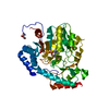
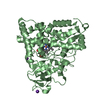
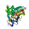
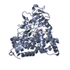
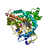
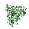
| Entry | Database: PDB / ID: 4n9q | ||||||
|---|---|---|---|---|---|---|---|
| Title | Crystal structure of paAzoR1 bound to ubiquinone-1 | ||||||
 Components Components | FMN-dependent NADH-azoreductase 1 | ||||||
 Keywords Keywords |  OXIDOREDUCTASE / azoreductase / quinone / NAD(P)H quinone oxidoreductase / ubiquinone-1 OXIDOREDUCTASE / azoreductase / quinone / NAD(P)H quinone oxidoreductase / ubiquinone-1 | ||||||
| Function / homology |  Function and homology informationNADPH:quinone reductase activity / Oxidoreductases; Acting on NADH or NADPH; With a quinone or similar compound as acceptor / FMN-dependent NADH-azoreductase / oxidoreductase activity, acting on NAD(P)H, NAD(P) as acceptor / oxidoreductase activity, acting on NAD(P)H, quinone or similar compound as acceptor / FMN binding / electron transfer activity Function and homology informationNADPH:quinone reductase activity / Oxidoreductases; Acting on NADH or NADPH; With a quinone or similar compound as acceptor / FMN-dependent NADH-azoreductase / oxidoreductase activity, acting on NAD(P)H, NAD(P) as acceptor / oxidoreductase activity, acting on NAD(P)H, quinone or similar compound as acceptor / FMN binding / electron transfer activitySimilarity search - Function | ||||||
| Biological species |   Pseudomonas aeruginosa (bacteria) Pseudomonas aeruginosa (bacteria) | ||||||
| Method | X-RAY DIFFRACTION / SYNCHROTRON / MOLECULAR REPLACEMENT / molecular replacement / Resolution: 2 Å | ||||||
 Authors Authors | Ryan, A. / Kaplan, E. / Crescente, V. / Lowe, E. / Preston, G.M. / Sim, E. | ||||||
 Citation Citation | Journal: Plos One / Year: 2014 Title: Identification of NAD(P)H Quinone Oxidoreductase Activity in Azoreductases from P. aeruginosa: Azoreductases and NAD(P)H Quinone Oxidoreductases Belong to the Same FMN-Dependent Superfamily of Enzymes. Authors: Ryan, A. / Kaplan, E. / Nebel, J.C. / Polycarpou, E. / Crescente, V. / Lowe, E. / Preston, G.M. / Sim, E. | ||||||
| History |
|
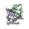
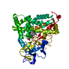
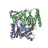
- Structure visualization
| Structure viewer | Molecule: MolmilJmol/JSmol |
|---|
- Downloads & links
-Download
| PDBx/mmCIF format | 4n9q.cif.gz | 230.4 KB | Display | PDBx/mmCIF format |
|---|---|---|---|---|
| PDB format | pdb4n9q.ent.gz | 187.1 KB | Display | PDB format |
| PDBx/mmJSON format | 4n9q.json.gz | Tree view | PDBx/mmJSON format | |
| Others |  Other downloads Other downloads |
-Validation report
| Arichive directory | https://data.pdbj.org/pub/pdb/validation_reports/n9/4n9qftp://data.pdbj.org/pub/pdb/validation_reports/n9/4n9q | HTTPS FTP |
|---|
-Related structure data
| Related structure data |  4n65C 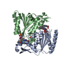 2v9cS S: Starting model for refinement C: citing same article ( |
|---|---|
| Similar structure data |


-Links
 PDBj
PDBj
- Assembly
| Deposited unit | 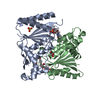
| ||||||||
|---|---|---|---|---|---|---|---|---|---|
| 1 |
| ||||||||
| Unit cell |
| ||||||||
| Components on special symmetry positions |
|
-Components
-Protein , 1 types, 2 molecules AB
| #1: Protein | Mass: 23077.074 Da / Num. of mol.: 2 Source method: isolated from a genetically manipulated source Source: (gene. exp.) Pseudomonas aeruginosa (bacteria) / Strain: ATCC 15692 /PAO1 /1C /PRS 101 /LMG 12228 / Gene: azoR1, PA0785 / Plasmid: pET28b / Production host: Escherichia coli (E. coli) / Strain (production host): BL21(DE3) plyssReferences: UniProt: Q9I5F3, Oxidoreductases; Acting on other nitrogenous compounds as donors |
|---|
-Non-polymers , 5 types, 161 molecules 
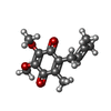


| #2: Chemical | Flavin mononucleotide Mass: 456.344 Da / Num. of mol.: 2 / Source method: obtained synthetically / Formula: C17H21N4O9P Mass: 456.344 Da / Num. of mol.: 2 / Source method: obtained synthetically / Formula: C17H21N4O9P#3: Chemical | ChemComp-UQ1 / |  Mass: 250.290 Da / Num. of mol.: 1 / Source method: obtained synthetically / Formula: C14H18O4 Mass: 250.290 Da / Num. of mol.: 1 / Source method: obtained synthetically / Formula: C14H18O4#4: Chemical | ChemComp-GOL / | Glycerol Mass: 92.094 Da / Num. of mol.: 1 / Source method: obtained synthetically / Formula: C3H8O3 Mass: 92.094 Da / Num. of mol.: 1 / Source method: obtained synthetically / Formula: C3H8O3#5: Chemical | ChemComp-SO4 / | Sulfate Mass: 96.063 Da / Num. of mol.: 1 / Source method: obtained synthetically / Formula: SO4 Mass: 96.063 Da / Num. of mol.: 1 / Source method: obtained synthetically / Formula: SO4#6: Water | ChemComp-HOH / | WaterMass: 18.015 Da / Num. of mol.: 156 / Source method: isolated from a natural source / Formula: H2O |
|---|
-Experimental details
-Experiment
| Experiment | Method: X-RAY DIFFRACTION / Number of used crystals: 1 |
|---|
- Sample preparation
| Crystal | Density Matthews: 2.3 Å3/Da / Density % sol: 46.6 % |
|---|---|
| Crystal grow | Temperature: 298 K / Method: vapor diffusion / pH: 7.5 Details: 1.6 M ammonium sulphate, 0.1 M HEPES pH 7.5, vapor diffusion, temperature 298K |
-Data collection
| Diffraction | Mean temperature: 100 K | ||||||||||||||||||||||||||||||||||||||||||||||||||||||||||||||||||||||||||||||||||||||||
|---|---|---|---|---|---|---|---|---|---|---|---|---|---|---|---|---|---|---|---|---|---|---|---|---|---|---|---|---|---|---|---|---|---|---|---|---|---|---|---|---|---|---|---|---|---|---|---|---|---|---|---|---|---|---|---|---|---|---|---|---|---|---|---|---|---|---|---|---|---|---|---|---|---|---|---|---|---|---|---|---|---|---|---|---|---|---|---|---|---|
| Diffraction source | Source: SYNCHROTRON / Site: Diamond  / Beamline: I03 / Wavelength: 0.8726 Å / Beamline: I03 / Wavelength: 0.8726 Å | ||||||||||||||||||||||||||||||||||||||||||||||||||||||||||||||||||||||||||||||||||||||||
| Detector | Type: ADSC QUANTUM 210r / Detector: CCD / Date: Dec 2, 2011 | ||||||||||||||||||||||||||||||||||||||||||||||||||||||||||||||||||||||||||||||||||||||||
| Radiation | Protocol: SINGLE WAVELENGTH / Scattering type: x-ray | ||||||||||||||||||||||||||||||||||||||||||||||||||||||||||||||||||||||||||||||||||||||||
| Radiation wavelength | Wavelength: 0.8726 Å / Relative weight: 1 | ||||||||||||||||||||||||||||||||||||||||||||||||||||||||||||||||||||||||||||||||||||||||
| Reflection | Resolution: 2→71.362 Å / Num. all: 26575 / Num. obs: 26575 / % possible obs: 90.7 % / Redundancy: 4.7 % / Rsym value: 0.053 / Net I/σ(I): 17.1 | ||||||||||||||||||||||||||||||||||||||||||||||||||||||||||||||||||||||||||||||||||||||||
| Reflection shell | Diffraction-ID: 1
|
-Phasing
| Phasing | Method: molecular replacement |
|---|
- Processing
| Software |
| |||||||||||||||||||||||||||||||||||||||||||||||||||||||||||||||||||||||||||||
|---|---|---|---|---|---|---|---|---|---|---|---|---|---|---|---|---|---|---|---|---|---|---|---|---|---|---|---|---|---|---|---|---|---|---|---|---|---|---|---|---|---|---|---|---|---|---|---|---|---|---|---|---|---|---|---|---|---|---|---|---|---|---|---|---|---|---|---|---|---|---|---|---|---|---|---|---|---|---|
| Refinement | Method to determine structure: MOLECULAR REPLACEMENT Starting model: PDB entry 2V9C Resolution: 2→43.18 Å / Occupancy max: 1 / Occupancy min: 0 / FOM work R set: 0.7707 / SU ML: 0.26 / σ(F): 1.36 / Phase error: 27.13 / Stereochemistry target values: ML
| |||||||||||||||||||||||||||||||||||||||||||||||||||||||||||||||||||||||||||||
| Solvent computation | Shrinkage radii: 0.9 Å / VDW probe radii: 1.11 Å / Solvent model: FLAT BULK SOLVENT MODEL | |||||||||||||||||||||||||||||||||||||||||||||||||||||||||||||||||||||||||||||
| Displacement parameters | Biso max: 128.56 Å2 / Biso mean: 49.2038 Å2 / Biso min: 20 Å2 | |||||||||||||||||||||||||||||||||||||||||||||||||||||||||||||||||||||||||||||
| Refinement step | Cycle: LAST / Resolution: 2→43.18 Å
| |||||||||||||||||||||||||||||||||||||||||||||||||||||||||||||||||||||||||||||
| Refine LS restraints |
| |||||||||||||||||||||||||||||||||||||||||||||||||||||||||||||||||||||||||||||
| LS refinement shell | Refine-ID: X-RAY DIFFRACTION / Total num. of bins used: 10
| |||||||||||||||||||||||||||||||||||||||||||||||||||||||||||||||||||||||||||||
| Refinement TLS params. | Method: refined / Origin x: 24.7322 Å / Origin y: -13.6114 Å / Origin z: 11.766 Å
| |||||||||||||||||||||||||||||||||||||||||||||||||||||||||||||||||||||||||||||
| Refinement TLS group |
|
