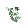
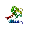
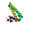
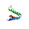
Entry Database : PDB / ID : 4myzTitle Structure of a class 2 docking domain complex from modules CurK and CurL of the curacin A polyketide synthase CurK, CurL fusion protein Keywords / / Function / homology Function Domain/homology Component
/ / / / / / / / / / / / / / / / / / / / / / / / / / / / / / / / / / / / / / / / / / / / / / / / / / / / / / / / / / / / / / / / / / / / / / Biological species Moorea producens 3L (bacteria)Method / / / Resolution : 1.5 Å Authors Whicher, J.R. / Smaga, S.S. / Smith, J.L. Journal : Chem.Biol. / Year : 2013Title : Cyanobacterial polyketide synthase docking domains: a tool for engineering natural product biosynthesis.Authors : Whicher, J.R. / Smaga, S.S. / Hansen, D.A. / Brown, W.C. / Gerwick, W.H. / Sherman, D.H. / Smith, J.L. History Deposition Sep 28, 2013 Deposition site / Processing site Revision 1.0 Jan 29, 2014 Provider / Type Revision 1.1 Feb 5, 2014 Group Revision 1.2 Aug 9, 2017 Group / Refinement description / Source and taxonomyCategory / entity_src_gen / software / Item Revision 1.3 Feb 28, 2024 Group / Database referencesCategory chem_comp_atom / chem_comp_bond ... chem_comp_atom / chem_comp_bond / database_2 / struct_ref_seq_dif Item / _database_2.pdbx_database_accession / _struct_ref_seq_dif.details
Show all Show less
 Movie
Movie Controller

 Yorodumi
 Open data
 Basic information
 Components
 Keywords
 PROTEIN BINDING /
  Function and homology information
 Moorea producens 3L (bacteria)
 Authors
 Citation
 Structure visualization
 Downloads & links
 Other downloads
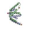


 PDBj
PDBj


 Assembly


 Mass: 18.015 Da / Num. of mol.: 160 / Source method: isolated from a natural source / Formula: H2O
 Sample preparation
 / Beamline: 23-ID-D / Wavelength: 1.033 Å
 Processing