 Movie
Movie Controller


+ Open data

- Basic information
| Entry | Database: PDB / ID: 2f05 | ||||||
|---|---|---|---|---|---|---|---|
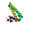
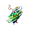
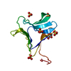
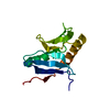
| Title | Solution structure of free PAH2 domain of mSin3B | ||||||
 Components Components | Paired amphipathic helix protein Sin3b | ||||||
 Keywords Keywords | TRANSCRIPTION REPRESSOR / 4 helix bundle | ||||||
| Function / homology |  Function and homology information Function and homology information autosome / RUNX1 regulates genes involved in megakaryocyte differentiation and platelet function / Regulation of lipid metabolism by PPARalpha / Cytoprotection by HMOX1 / XY body / cardiac muscle tissue development / Sin3-type complex / Y chromosome / X chromosome / negative regulation of cell cycle ...autosome / RUNX1 regulates genes involved in megakaryocyte differentiation and platelet function / Regulation of lipid metabolism by PPARalpha / Cytoprotection by HMOX1 / XY body / cardiac muscle tissue development / Sin3-type complex / Y chromosome / X chromosome / negative regulation of cell cycle / skeletal muscle tissue development / negative regulation of cell migration / transcription corepressor activity / negative regulation of DNA-templated transcription / chromatin binding / chromatin / negative regulation of transcription by RNA polymerase II / nucleus / cytoplasm autosome / RUNX1 regulates genes involved in megakaryocyte differentiation and platelet function / Regulation of lipid metabolism by PPARalpha / Cytoprotection by HMOX1 / XY body / cardiac muscle tissue development / Sin3-type complex / Y chromosome / X chromosome / negative regulation of cell cycle ...autosome / RUNX1 regulates genes involved in megakaryocyte differentiation and platelet function / Regulation of lipid metabolism by PPARalpha / Cytoprotection by HMOX1 / XY body / cardiac muscle tissue development / Sin3-type complex / Y chromosome / X chromosome / negative regulation of cell cycle / skeletal muscle tissue development / negative regulation of cell migration / transcription corepressor activity / negative regulation of DNA-templated transcription / chromatin binding / chromatin / negative regulation of transcription by RNA polymerase II / nucleus / cytoplasmSimilarity search - Function | ||||||
| Biological species |  Mus musculus (house mouse) Mus musculus (house mouse) | ||||||
| Method | SOLUTION NMR / simulated annealing, with water-refinement | ||||||
 Authors Authors | van Ingen, H. / Baltussen, M.A. / Aelen, J. / Vuister, G.W. | ||||||
 Citation Citation | Journal: J.Mol.Biol. / Year: 2006 Title: Role of Structural and Dynamical Plasticity in Sin3: The Free PAH2 Domain is a Folded Module in mSin3B Authors: van Ingen, H. / Baltussen, M.A. / Aelen, J. / Vuister, G.W. | ||||||
| History |
|
- Structure visualization
| Structure viewer | Molecule: MolmilJmol/JSmol |
|---|
- Downloads & links
-Download
| PDBx/mmCIF format | 2f05.cif.gz | 927 KB | Display | PDBx/mmCIF format |
|---|---|---|---|---|
| PDB format | pdb2f05.ent.gz | 792.5 KB | Display | PDB format |
| PDBx/mmJSON format | 2f05.json.gz | Tree view | PDBx/mmJSON format | |
| Others |  Other downloads Other downloads |
-Validation report
| Arichive directory | https://data.pdbj.org/pub/pdb/validation_reports/f0/2f05ftp://data.pdbj.org/pub/pdb/validation_reports/f0/2f05 | HTTPS FTP |
|---|
-Related structure data
| Related structure data | |
|---|---|
| Similar structure data | |
| Other databases |
|


-Links
 PDBj
PDBj


- Assembly
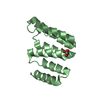
| Deposited unit | 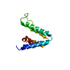
| |||||||||
|---|---|---|---|---|---|---|---|---|---|---|
| 1 |
| |||||||||
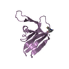
| NMR ensembles |
|
-Components
| #1: Protein | Mass: 12241.564 Da / Num. of mol.: 1 / Fragment: PAH2 domain (residues 148-252) Source method: isolated from a genetically manipulated source Source: (gene. exp.) Mus musculus (house mouse) / Gene: Sin3b, Kiaa0700 / Plasmid: pGEX2T / Species (production host): Escherichia coli / Production host:  Escherichia coli BL21(DE3) (bacteria) / Strain (production host): BL21-DE3 / References: UniProt: Q62141 Escherichia coli BL21(DE3) (bacteria) / Strain (production host): BL21-DE3 / References: UniProt: Q62141 |
|---|
-Experimental details
-Experiment
| Experiment | Method: SOLUTION NMR |
|---|
- Sample preparation
| Details | Contents: 0.9 mM PAH2 uniform 13C/15N labeled, H2O, 100mM KCl, 50 mM Pi buffer pH 6.3 Solvent system: H2O, 100mM KCl, 50 mM Pi buffer pH 6.3 |
|---|---|
| Sample conditions | Ionic strength: 150 / pH: 6.3 / Pressure: 1 atm / Temperature: 293 K |
-NMR measurement
| Radiation | Protocol: SINGLE WAVELENGTH / Monochromatic (M) / Laue (L): M |
|---|---|
| Radiation wavelength | Relative weight: 1 |
| NMR spectrometer | Type: Varian UNITY / Manufacturer: Varian / Model: UNITY / Field strength: 600 MHz |
- Processing
| NMR software |
| ||||||||||||
|---|---|---|---|---|---|---|---|---|---|---|---|---|---|
| Refinement | Method: simulated annealing, with water-refinement / Software ordinal: 1 | ||||||||||||
| NMR representative | Selection criteria: closest to the average | ||||||||||||
| NMR ensemble | Conformer selection criteria: structures with the lowest energy Conformers calculated total number: 200 / Conformers submitted total number: 30 |
