 Movie
Movie Controller

[English] 日本語
 Yorodumi
- PDB-4ml3: X-ray structure of ComE D58A REC domain from Streptococcus pneumoniae -
+ Open data

- Basic information
| Entry | Database: PDB / ID: 4ml3 | ||||||
|---|---|---|---|---|---|---|---|
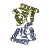
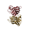
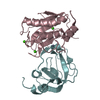
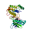
| Title | X-ray structure of ComE D58A REC domain from Streptococcus pneumoniae | ||||||
 Components Components | Response regulator | ||||||
 Keywords Keywords | UNKNOWN FUNCTION / protein Dimer / REC / response regulator | ||||||
| Function / homology |  Function and homology information Function and homology information | ||||||
| Biological species |   Streptococcus pneumoniae (bacteria) Streptococcus pneumoniae (bacteria) | ||||||
| Method | X-RAY DIFFRACTION / SYNCHROTRON / MOLECULAR REPLACEMENT / Resolution: 3.15 Å | ||||||
 Authors Authors | Boudes, M. / Sanchez, D. / Durand, D. / Graille, M. / van Tilbeurgh, H. / Quevillon-Cheruel, S. | ||||||
 Citation Citation | Journal: Nucleic Acids Res. / Year: 2014 Title: Structural insights into the dimerization of the response regulator ComE from Streptococcus pneumoniae. Authors: Boudes, M. / Sanchez, D. / Graille, M. / van Tilbeurgh, H. / Durand, D. / Quevillon-Cheruel, S. | ||||||
| History |
|
- Structure visualization
| Structure viewer | Molecule: MolmilJmol/JSmol |
|---|
- Downloads & links
-Download
| PDBx/mmCIF format | 4ml3.cif.gz | 114.6 KB | Display | PDBx/mmCIF format |
|---|---|---|---|---|
| PDB format | pdb4ml3.ent.gz | 91.4 KB | Display | PDB format |
| PDBx/mmJSON format | 4ml3.json.gz | Tree view | PDBx/mmJSON format | |
| Others |  Other downloads Other downloads |
-Validation report
| Arichive directory | https://data.pdbj.org/pub/pdb/validation_reports/ml/4ml3ftp://data.pdbj.org/pub/pdb/validation_reports/ml/4ml3 | HTTPS FTP |
|---|
-Related structure data
| Related structure data |  4cbvC  4mldC  4b1n S: Starting model for refinement C: citing same article ( |
|---|---|
| Similar structure data |


-Links
 PDBj
PDBj

- Assembly
| Deposited unit | 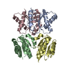
| ||||||||
|---|---|---|---|---|---|---|---|---|---|
| 1 | 
| ||||||||
| 2 | 
| ||||||||
| Unit cell |
|
-Components
| #1: Protein | Mass: 17169.479 Da / Num. of mol.: 4 / Fragment: REC domain (UNP residues 1-137) / Mutation: D58A Source method: isolated from a genetically manipulated source Source: (gene. exp.) Streptococcus pneumoniae (bacteria) / Strain: ATCC BAA-255 / R6 / Gene: comE, spr2041 / Plasmid: pET28a / Production host: Escherichia coli (E. coli) / Strain (production host): BL21-Gold (DE3) / References: UniProt: Q8DMW5 |
|---|
-Experimental details
-Experiment
| Experiment | Method: X-RAY DIFFRACTION / Number of used crystals: 1 |
|---|
- Sample preparation
| Crystal | Density Matthews: 2.37 Å3/Da / Density % sol: 48.12 % |
|---|---|
| Crystal grow | Temperature: 291.2 K / Method: vapor diffusion, hanging drop / pH: 7.5 Details: 25% PEG 3350, 0.2M Sodium formate , pH 7.5, VAPOR DIFFUSION, HANGING DROP, temperature 291.2K |
-Data collection
| Diffraction | Mean temperature: 100 K |
|---|---|
| Diffraction source | Source: SYNCHROTRON / Site: SOLEIL  / Beamline: PROXIMA 1 / Wavelength: 0.98011 Å / Beamline: PROXIMA 1 / Wavelength: 0.98011 Å |
| Detector | Type: PSI PILATUS 6M / Detector: PIXEL / Date: Feb 24, 2013 |
| Radiation | Monochromator: channel cut cryogenically cooled monochromator crystal Protocol: SINGLE WAVELENGTH / Monochromatic (M) / Laue (L): M / Scattering type: x-ray |
| Radiation wavelength | Wavelength: 0.98011 Å / Relative weight: 1 |
| Reflection | Resolution: 3.15→45.7 Å / Num. all: 11136 / Num. obs: 11099 / % possible obs: 99.67 % / Observed criterion σ(F): 3.16 / Observed criterion σ(I): 3.16 / Redundancy: 9.22 % / Biso Wilson estimate: 65.55 Å2 / Rmerge(I) obs: 0.144 / Rsym value: 0.144 / Net I/σ(I): 15 |
| Reflection shell | Resolution: 3.15→3.46 Å / Redundancy: 9.2 % / Rmerge(I) obs: 0.144 / Mean I/σ(I) obs: 15 / Num. unique all: 11099 / Rsym value: 0.144 / % possible all: 99.7 |
- Processing
| Software |
| |||||||||||||||||||||||||||||||||||
|---|---|---|---|---|---|---|---|---|---|---|---|---|---|---|---|---|---|---|---|---|---|---|---|---|---|---|---|---|---|---|---|---|---|---|---|---|
| Refinement | Method to determine structure: MOLECULAR REPLACEMENT Starting model: PDB entry 4B1N 4b1n Resolution: 3.15→45.7 Å / SU ML: 0.15 / σ(F): 1.99 / σ(I): 2 / Phase error: 33.2 / Stereochemistry target values: LS_WUNIT_K1
| |||||||||||||||||||||||||||||||||||
| Solvent computation | Shrinkage radii: 0.86 Å / VDW probe radii: 1.1 Å / Solvent model: FLAT BULK SOLVENT MODEL / Bsol: 69.909 Å2 / ksol: 0.14 e/Å3 | |||||||||||||||||||||||||||||||||||
| Displacement parameters |
| |||||||||||||||||||||||||||||||||||
| Refinement step | Cycle: LAST / Resolution: 3.15→45.7 Å
| |||||||||||||||||||||||||||||||||||
| Refine LS restraints |
| |||||||||||||||||||||||||||||||||||
| LS refinement shell |
|
