 Movie
Movie Controller

[English] 日本語
 Yorodumi
- PDB-4mej: Crystal structure of Lactobacillus helveticus purine deoxyribosyl... -
+ Open data

- Basic information
| Entry | Database: PDB / ID: 4mej | ||||||
|---|---|---|---|---|---|---|---|
| Title | Crystal structure of Lactobacillus helveticus purine deoxyribosyl transferase (PDT) with the tricyclic purine 8,9-dihydro-9-oxoimidazo[2,1-b]purine (N2,3-ethenoguanine) | ||||||
 Components Components | Nucleoside deoxyribosyltransferase | ||||||
 Keywords Keywords | transferase/transferase inhibitor / purine deoxyribosyl transferase (PDT) / transferase-transferase inhibitor complex | ||||||
| Function / homology | Nucleoside 2-deoxyribosyltransferase / Nucleoside 2-deoxyribosyltransferase / Rossmann fold - #450 / transferase activity / Rossmann fold / 3-Layer(aba) Sandwich / Alpha Beta / 3H-imidazo[2,1-b]purin-4(5H)-one / Nucleoside deoxyribosyltransferase Function and homology information Function and homology information | ||||||
| Biological species |  Lactobacillus helveticus (bacteria) Lactobacillus helveticus (bacteria) | ||||||
| Method | X-RAY DIFFRACTION / SYNCHROTRON / MOLECULAR REPLACEMENT / Resolution: 2.1 Å | ||||||
 Authors Authors | Paul, D. / Seckute, J. / Ealick, S.E. | ||||||
 Citation Citation | Journal: Plos One / Year: 2014 Title: Glycosylation of a tricyclic purine analog at alternative sites by nucleoside 2 -deoxyribosyltransferases Authors: Ye, W. / Paul, D. / Gao, L. / Seckute, J. / Sangaiah, R. / Karupiah, J. / Zhang, Z. / Kaminski, A.P. / Ealick, S.E. / Gold, A. / Ball, L.M. | ||||||
| History |
|
- Structure visualization
| Structure viewer | Molecule: MolmilJmol/JSmol |
|---|
- Downloads & links
-Download
| PDBx/mmCIF format | 4mej.cif.gz | 117.3 KB | Display | PDBx/mmCIF format |
|---|---|---|---|---|
| PDB format | pdb4mej.ent.gz | 91.7 KB | Display | PDB format |
| PDBx/mmJSON format | 4mej.json.gz | Tree view | PDBx/mmJSON format | |
| Others |  Other downloads Other downloads |
-Validation report
| Arichive directory | https://data.pdbj.org/pub/pdb/validation_reports/me/4mejftp://data.pdbj.org/pub/pdb/validation_reports/me/4mej | HTTPS FTP |
|---|
-Related structure data
| Related structure data |  1s2lS S: Starting model for refinement |
|---|---|
| Similar structure data |


-Links
 PDBj
PDBj- Assembly
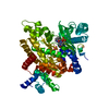
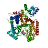
| Deposited unit | 
| ||||||||
|---|---|---|---|---|---|---|---|---|---|
| 1 |
| ||||||||
| Unit cell |
| ||||||||
| Noncrystallographic symmetry (NCS) | NCS domain:
NCS domain segments: (Selection details: chain 'C' RESTRAINED TORSIONS: 2342 Histogram of differences under limit:...) |
-Components
| #1: Protein | Mass: 18733.189 Da / Num. of mol.: 3 Source method: isolated from a genetically manipulated source Source: (gene. exp.) Lactobacillus helveticus (bacteria) / Strain: H10 / Gene: LBHH_1694 / Production host: Escherichia coli (E. coli) / References: UniProt: F0NW64#2: Chemical | Sulfate  Mass: 96.063 Da / Num. of mol.: 2 / Source method: obtained synthetically / Formula: SO4 Mass: 96.063 Da / Num. of mol.: 2 / Source method: obtained synthetically / Formula: SO4#3: Chemical |   Mass: 175.147 Da / Num. of mol.: 3 / Source method: obtained synthetically / Formula: C7H5N5O Mass: 175.147 Da / Num. of mol.: 3 / Source method: obtained synthetically / Formula: C7H5N5O#4: Water | ChemComp-HOH / | Water Mass: 18.015 Da / Num. of mol.: 282 / Source method: isolated from a natural source / Formula: H2O Mass: 18.015 Da / Num. of mol.: 282 / Source method: isolated from a natural source / Formula: H2O |
|---|
-Experimental details
-Experiment
| Experiment | Method: X-RAY DIFFRACTION / Number of used crystals: 1 |
|---|
- Sample preparation
| Crystal | Density Matthews: 2.64 Å3/Da / Density % sol: 53.36 % |
|---|---|
| Crystal grow | Temperature: 295 K / Method: vapor diffusion, hanging drop / pH: 7.9 Details: 100 mM Tris and 2.2 M ammonium sulfate, pH 7.9, VAPOR DIFFUSION, HANGING DROP, temperature 295K |
-Data collection
| Diffraction | Mean temperature: 80 K |
|---|---|
| Diffraction source | Source: SYNCHROTRON / Site: APS  / Beamline: 24-ID-E / Wavelength: 0.98 Å / Beamline: 24-ID-E / Wavelength: 0.98 Å |
| Detector | Type: ADSC QUANTUM 315 / Detector: CCD / Date: Oct 1, 2007 |
| Radiation | Monochromator: Cryogenically-cooled single crystal Si(111) side bounce monochromator Protocol: SINGLE WAVELENGTH / Monochromatic (M) / Laue (L): M / Scattering type: x-ray |
| Radiation wavelength | Wavelength: 0.98 Å / Relative weight: 1 |
| Reflection | Resolution: 2.1→50 Å / Num. all: 35578 / Num. obs: 35578 / % possible obs: 100 % / Observed criterion σ(F): 1 / Observed criterion σ(I): 1 / Redundancy: 5.9 % / Rmerge(I) obs: 0.049 / Rsym value: 0.046 / Net I/σ(I): 23.1 |
| Reflection shell | Resolution: 2.1→2.18 Å / Redundancy: 5.3 % / Rmerge(I) obs: 0.235 / Mean I/σ(I) obs: 4.8 / Num. unique all: 3279 / Rsym value: 0.266 / % possible all: 100 |
- Processing
| Software |
| ||||||||||||||||||||||||||||||||||||||||||||||||||||||||||||||||||||||||||||||||||||||||||||||||||
|---|---|---|---|---|---|---|---|---|---|---|---|---|---|---|---|---|---|---|---|---|---|---|---|---|---|---|---|---|---|---|---|---|---|---|---|---|---|---|---|---|---|---|---|---|---|---|---|---|---|---|---|---|---|---|---|---|---|---|---|---|---|---|---|---|---|---|---|---|---|---|---|---|---|---|---|---|---|---|---|---|---|---|---|---|---|---|---|---|---|---|---|---|---|---|---|---|---|---|---|
| Refinement | Method to determine structure: MOLECULAR REPLACEMENT Starting model: pdb entry 1S2L Resolution: 2.1→36.648 Å / SU ML: 0.21 / σ(F): 1.34 / Phase error: 22.54 / Stereochemistry target values: ML
| ||||||||||||||||||||||||||||||||||||||||||||||||||||||||||||||||||||||||||||||||||||||||||||||||||
| Solvent computation | Shrinkage radii: 0.9 Å / VDW probe radii: 1.11 Å / Solvent model: FLAT BULK SOLVENT MODEL | ||||||||||||||||||||||||||||||||||||||||||||||||||||||||||||||||||||||||||||||||||||||||||||||||||
| Refinement step | Cycle: LAST / Resolution: 2.1→36.648 Å
| ||||||||||||||||||||||||||||||||||||||||||||||||||||||||||||||||||||||||||||||||||||||||||||||||||
| Refine LS restraints |
| ||||||||||||||||||||||||||||||||||||||||||||||||||||||||||||||||||||||||||||||||||||||||||||||||||
| LS refinement shell |
|
