 Movie
Movie Controller


+ Open data

- Basic information
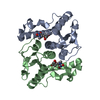
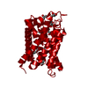
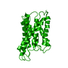
| Entry | Database: PDB / ID: 1s2g | ||||||
|---|---|---|---|---|---|---|---|
| Title | Purine 2'deoxyribosyltransferase + 2'-deoxyadenosine | ||||||
 Components Components | purine trans deoxyribosylase | ||||||
 Keywords Keywords |  TRANSFERASE / PTD / 2'-deoxyadenosine / 2'-purine deoxyribosyltansferase TRANSFERASE / PTD / 2'-deoxyadenosine / 2'-purine deoxyribosyltansferase | ||||||
| Function / homology |  Function and homology informationnucleoside deoxyribosyltransferase / nucleoside deoxyribosyltransferase activity Function and homology informationnucleoside deoxyribosyltransferase / nucleoside deoxyribosyltransferase activitySimilarity search - Function | ||||||
| Biological species |  Lactobacillus helveticus (bacteria) Lactobacillus helveticus (bacteria) | ||||||
| Method | X-RAY DIFFRACTION / SYNCHROTRON / MOLECULAR REPLACEMENT / Resolution: 2.1 Å | ||||||
 Authors Authors | Anand, R. / Kaminski, P.A. / Ealick, S.E. | ||||||
 Citation Citation | Journal: Biochemistry / Year: 2004 Title: Structures of purine 2'-deoxyribosyltransferase, substrate complexes, and the ribosylated enzyme intermediate at 2.0 A resolution. Authors: Anand, R. / Kaminski, P.A. / Ealick, S.E. | ||||||
| History |
|
- Structure visualization
| Structure viewer | Molecule: MolmilJmol/JSmol |
|---|
- Downloads & links
-Download
| PDBx/mmCIF format | 1s2g.cif.gz | 105.6 KB | Display | PDBx/mmCIF format |
|---|---|---|---|---|
| PDB format | pdb1s2g.ent.gz | 82 KB | Display | PDB format |
| PDBx/mmJSON format | 1s2g.json.gz | Tree view | PDBx/mmJSON format | |
| Others |  Other downloads Other downloads |
-Validation report
| Arichive directory | https://data.pdbj.org/pub/pdb/validation_reports/s2/1s2gftp://data.pdbj.org/pub/pdb/validation_reports/s2/1s2g | HTTPS FTP |
|---|
-Related structure data


-Links
 PDBj
PDBj- Assembly
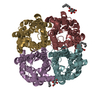
| Deposited unit | 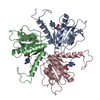
| ||||||||
|---|---|---|---|---|---|---|---|---|---|
| 1 | 
| ||||||||
| Unit cell |
|
-Components
| #1: Protein | Mass: 18733.189 Da / Num. of mol.: 3 / Fragment: purine 2'-deoxyribosyltansferase Source method: isolated from a genetically manipulated source Source: (gene. exp.) Lactobacillus helveticus (bacteria) / Production host: Escherichia coli (E. coli)References: UniProt: Q8RLY5, nucleoside deoxyribosyltransferase#2: Chemical | Deoxyadenosine  Mass: 251.242 Da / Num. of mol.: 3 / Source method: obtained synthetically / Formula: C10H13N5O3 Mass: 251.242 Da / Num. of mol.: 3 / Source method: obtained synthetically / Formula: C10H13N5O3#3: Water | ChemComp-HOH / | Water Mass: 18.015 Da / Num. of mol.: 272 / Source method: isolated from a natural source / Formula: H2O Mass: 18.015 Da / Num. of mol.: 272 / Source method: isolated from a natural source / Formula: H2O |
|---|
-Experimental details
-Experiment
| Experiment | Method: X-RAY DIFFRACTION / Number of used crystals: 1 |
|---|
- Sample preparation
| Crystal | Density Matthews: 2.71 Å3/Da / Density % sol: 54.18 % |
|---|---|
| Crystal grow | Temperature: 295 K / Method: vapor diffusion, hanging drop / pH: 8 Details: 2.0 M ammonium sulfate, 100mM Tris , pH 8.0, VAPOR DIFFUSION, HANGING DROP, temperature 295K |
-Data collection
| Diffraction | Mean temperature: 170 K |
|---|---|
| Diffraction source | Source: SYNCHROTRON / Site: APS  / Beamline: 8-BM / Wavelength: 0.9791 Å / Beamline: 8-BM / Wavelength: 0.9791 Å |
| Detector | Type: ADSC QUANTUM 315 / Detector: CCD / Date: Sep 18, 2002 |
| Radiation | Protocol: SINGLE WAVELENGTH / Monochromatic (M) / Laue (L): M / Scattering type: x-ray |
| Radiation wavelength | Wavelength: 0.9791 Å / Relative weight: 1 |
| Reflection | Resolution: 2.1→25 Å / Num. all: 40463 / Num. obs: 40463 / % possible obs: 97.6 % / Observed criterion σ(F): 0 / Observed criterion σ(I): 0 / Redundancy: 8 % / Biso Wilson estimate: 19.4 Å2 / Rmerge(I) obs: 0.066 / Rsym value: 0.068 / Net I/σ(I): 20 |
| Reflection shell | Resolution: 2.1→2.2 Å / Redundancy: 8 % / Rmerge(I) obs: 0.334 / Mean I/σ(I) obs: 10.1 / Rsym value: 0.334 / % possible all: 99.8 |
- Processing
| Software |
| |||||||||||||||||||||||||
|---|---|---|---|---|---|---|---|---|---|---|---|---|---|---|---|---|---|---|---|---|---|---|---|---|---|---|
| Refinement | Method to determine structure: MOLECULAR REPLACEMENT / Resolution: 2.1→19.92 Å / Rfactor Rfree error: 0.005 / Data cutoff high absF: 265301.55 / Data cutoff low absF: 0 / Isotropic thermal model: RESTRAINED / Cross valid method: THROUGHOUT / σ(F): 0 / σ(I): 0 / Stereochemistry target values: Engh & Huber
| |||||||||||||||||||||||||
| Solvent computation | Solvent model: FLAT MODEL / Bsol: 33.0203 Å2 / ksol: 0.352912 e/Å3 | |||||||||||||||||||||||||
| Displacement parameters | Biso mean: 32.2 Å2
| |||||||||||||||||||||||||
| Refine analyze |
| |||||||||||||||||||||||||
| Refinement step | Cycle: LAST / Resolution: 2.1→19.92 Å
| |||||||||||||||||||||||||
| Refine LS restraints |
| |||||||||||||||||||||||||
| LS refinement shell | Resolution: 2.1→2.23 Å / Rfactor Rfree error: 0.014 / Total num. of bins used: 6
| |||||||||||||||||||||||||
| Xplor file |
|
