 Movie
Movie Controller


+ Open data

- Basic information
| Entry | Database: PDB / ID: 4mbb | ||||||
|---|---|---|---|---|---|---|---|
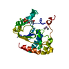
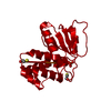
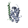
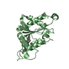
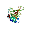
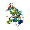
| Title | Cubic crystal form of PIR1 dual specificity phosphatase core | ||||||
 Components Components | RNA/RNP complex-1-interacting phosphatase | ||||||
 Keywords Keywords |  HYDROLASE / Atypical Dual Specificity Phosphatase / PIR1-core / RNA splicing / Helical hairpin / PTP-loop / deep catalytic cleft / Phosphate-binding loop (P-loop) / Acidic loop (WPD-loop) / RNA phosphatase / Dual specificity phosphatase / RNA-RNP complex-1 / dephosphorylation / nucleus HYDROLASE / Atypical Dual Specificity Phosphatase / PIR1-core / RNA splicing / Helical hairpin / PTP-loop / deep catalytic cleft / Phosphate-binding loop (P-loop) / Acidic loop (WPD-loop) / RNA phosphatase / Dual specificity phosphatase / RNA-RNP complex-1 / dephosphorylation / nucleus | ||||||
| Function / homology |  Function and homology information Function and homology informationprotein tyrosine/serine/threonine phosphatase activity / Hydrolases; Acting on ester bonds; Phosphoric-monoester hydrolases / polynucleotide 5'-phosphatase activity / intercellular bridge / phosphatase activity / RNA processing / protein dephosphorylation / protein tyrosine phosphatase activity / fibrillar center / nuclear speck ...protein tyrosine/serine/threonine phosphatase activity / Hydrolases; Acting on ester bonds; Phosphoric-monoester hydrolases / polynucleotide 5'-phosphatase activity / intercellular bridge / phosphatase activity / RNA processing / protein dephosphorylation / protein tyrosine phosphatase activity / fibrillar center / nuclear speck / RNA binding / nucleoplasm / nucleusSimilarity search - Function | ||||||
| Biological species |  Homo sapiens (human) Homo sapiens (human) | ||||||
| Method | X-RAY DIFFRACTION / SYNCHROTRON / MOLECULAR REPLACEMENT / Resolution: 1.849 Å | ||||||
 Authors Authors | Sankhala, R.S. / Lokareddy, R.K. / Cingolani, G. | ||||||
 Citation Citation | Journal: Biochemistry / Year: 2014 Title: Structure of Human PIR1, an Atypical Dual-Specificity Phosphatase. Authors: Sankhala, R.S. / Lokareddy, R.K. / Cingolani, G. | ||||||
| History |
|
- Structure visualization
| Structure viewer | Molecule: MolmilJmol/JSmol |
|---|
- Downloads & links
-Download
| PDBx/mmCIF format | 4mbb.cif.gz | 96.2 KB | Display | PDBx/mmCIF format |
|---|---|---|---|---|
| PDB format | pdb4mbb.ent.gz | 73.8 KB | Display | PDB format |
| PDBx/mmJSON format | 4mbb.json.gz | Tree view | PDBx/mmJSON format | |
| Others |  Other downloads Other downloads |
-Validation report
| Arichive directory | https://data.pdbj.org/pub/pdb/validation_reports/mb/4mbbftp://data.pdbj.org/pub/pdb/validation_reports/mb/4mbb | HTTPS FTP |
|---|
-Related structure data
| Related structure data | 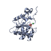 4nyhC  1yn9S S: Starting model for refinement C: citing same article ( |
|---|---|
| Similar structure data |


-Links
 PDBj
PDBj


- Assembly
| Deposited unit | 
| |||||||||
|---|---|---|---|---|---|---|---|---|---|---|
| 1 |
| |||||||||
| 2 | 
| |||||||||
| Unit cell |
| |||||||||
| Components on special symmetry positions |
|
-Components
| #1: Protein | Mass: 21477.498 Da / Num. of mol.: 1 / Fragment: UNP residues 29-207 / Mutation: C152S Source method: isolated from a genetically manipulated source Details: cell culture / Source: (gene. exp.) Homo sapiens (human) / Gene: DUSP11, PIR1 / Plasmid: pGEX-6P-1 / Production host:  Escherichia coli (E. coli) / Strain (production host): BL21(DE3) Escherichia coli (E. coli) / Strain (production host): BL21(DE3)References: UniProt: O75319, Hydrolases; Acting on ester bonds; Phosphoric-monoester hydrolases |
|---|---|
| #2: Chemical | ChemComp-CL / Chloride  Mass: 35.453 Da / Num. of mol.: 1 / Source method: obtained synthetically / Formula: Cl Mass: 35.453 Da / Num. of mol.: 1 / Source method: obtained synthetically / Formula: Cl |
| #3: Chemical | ChemComp-PO4 / Phosphate  Mass: 94.971 Da / Num. of mol.: 1 / Source method: obtained synthetically / Formula: PO4 Mass: 94.971 Da / Num. of mol.: 1 / Source method: obtained synthetically / Formula: PO4 |
| #4: Water | ChemComp-HOH / Water Mass: 18.015 Da / Num. of mol.: 181 / Source method: isolated from a natural source / Formula: H2O Mass: 18.015 Da / Num. of mol.: 181 / Source method: isolated from a natural source / Formula: H2O |
-Experimental details
-Experiment
| Experiment | Method: X-RAY DIFFRACTION / Number of used crystals: 1 |
|---|
- Sample preparation
| Crystal | Density Matthews: 3.09 Å3/Da / Density % sol: 60.26 % |
|---|---|
| Crystal grow | Temperature: 290 K / Method: vapor diffusion, hanging drop / pH: 5.5 Details: Bis-Tris 0.1M, NaCl 0.2M, polyethylene glycol 3350 22%, pH 5.5, VAPOR DIFFUSION, HANGING DROP, temperature 290K |
-Data collection
| Diffraction |
| |||||||||||||||
|---|---|---|---|---|---|---|---|---|---|---|---|---|---|---|---|---|
| Diffraction source |
| |||||||||||||||
| Detector |
| |||||||||||||||
| Radiation | Monochromator: Si 111 CHANNEL / Protocol: SINGLE WAVELENGTH / Monochromatic (M) / Laue (L): M / Scattering type: x-ray | |||||||||||||||
| Radiation wavelength |
| |||||||||||||||
| Reflection | Resolution: 1.85→15 Å / Num. obs: 22788 / % possible obs: 99.09 % / Observed criterion σ(F): 0 / Observed criterion σ(I): 0 | |||||||||||||||
| Reflection shell | Resolution: 1.85→1.92 Å / % possible all: 100 |

- Processing
| Software |
| |||||||||||||||||||||||||||||||||||||||||||||||||||||||||||||||||||||||||||||||||||||||||||||||||||||||||||||||||||||||||||||||||||||||||||||||||||||||||||||||||||||||||||||||||||||||||||||||||||||||||||||||||||||||||||||||||
|---|---|---|---|---|---|---|---|---|---|---|---|---|---|---|---|---|---|---|---|---|---|---|---|---|---|---|---|---|---|---|---|---|---|---|---|---|---|---|---|---|---|---|---|---|---|---|---|---|---|---|---|---|---|---|---|---|---|---|---|---|---|---|---|---|---|---|---|---|---|---|---|---|---|---|---|---|---|---|---|---|---|---|---|---|---|---|---|---|---|---|---|---|---|---|---|---|---|---|---|---|---|---|---|---|---|---|---|---|---|---|---|---|---|---|---|---|---|---|---|---|---|---|---|---|---|---|---|---|---|---|---|---|---|---|---|---|---|---|---|---|---|---|---|---|---|---|---|---|---|---|---|---|---|---|---|---|---|---|---|---|---|---|---|---|---|---|---|---|---|---|---|---|---|---|---|---|---|---|---|---|---|---|---|---|---|---|---|---|---|---|---|---|---|---|---|---|---|---|---|---|---|---|---|---|---|---|---|---|---|---|---|---|---|---|---|---|---|---|---|---|---|---|---|---|---|---|
| Refinement | Method to determine structure: MOLECULAR REPLACEMENT Starting model: PDB ENTRY 1YN9 Resolution: 1.849→14.664 Å / SU ML: 0.19 / σ(F): 0.82 / Phase error: 18.91 / Stereochemistry target values: ML
| |||||||||||||||||||||||||||||||||||||||||||||||||||||||||||||||||||||||||||||||||||||||||||||||||||||||||||||||||||||||||||||||||||||||||||||||||||||||||||||||||||||||||||||||||||||||||||||||||||||||||||||||||||||||||||||||||
| Solvent computation | Shrinkage radii: 0.9 Å / VDW probe radii: 1.11 Å / Solvent model: FLAT BULK SOLVENT MODEL | |||||||||||||||||||||||||||||||||||||||||||||||||||||||||||||||||||||||||||||||||||||||||||||||||||||||||||||||||||||||||||||||||||||||||||||||||||||||||||||||||||||||||||||||||||||||||||||||||||||||||||||||||||||||||||||||||
| Refinement step | Cycle: LAST / Resolution: 1.849→14.664 Å
| |||||||||||||||||||||||||||||||||||||||||||||||||||||||||||||||||||||||||||||||||||||||||||||||||||||||||||||||||||||||||||||||||||||||||||||||||||||||||||||||||||||||||||||||||||||||||||||||||||||||||||||||||||||||||||||||||
| Refine LS restraints |
| |||||||||||||||||||||||||||||||||||||||||||||||||||||||||||||||||||||||||||||||||||||||||||||||||||||||||||||||||||||||||||||||||||||||||||||||||||||||||||||||||||||||||||||||||||||||||||||||||||||||||||||||||||||||||||||||||
| LS refinement shell |
| |||||||||||||||||||||||||||||||||||||||||||||||||||||||||||||||||||||||||||||||||||||||||||||||||||||||||||||||||||||||||||||||||||||||||||||||||||||||||||||||||||||||||||||||||||||||||||||||||||||||||||||||||||||||||||||||||
| Refinement TLS params. | Method: refined / Refine-ID: X-RAY DIFFRACTION
| |||||||||||||||||||||||||||||||||||||||||||||||||||||||||||||||||||||||||||||||||||||||||||||||||||||||||||||||||||||||||||||||||||||||||||||||||||||||||||||||||||||||||||||||||||||||||||||||||||||||||||||||||||||||||||||||||
| Refinement TLS group |
|
