 Movie
Movie Controller


+ Open data

- Basic information
| Entry | Database: PDB / ID: 4m29 | ||||||
|---|---|---|---|---|---|---|---|
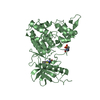
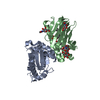
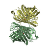
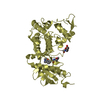
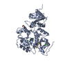
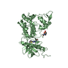
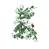
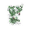
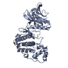
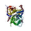
| Title | Structure of a GH39 Beta-xylosidase from Caulobacter crescentus | ||||||
 Components Components | Beta-xylosidase Xylan 1,4-b-xylosidase Xylan 1,4-b-xylosidase | ||||||
 Keywords Keywords | HYDROLASE / family GH39 / beta-xylosidase | ||||||
| Function / homology |  Function and homology information Function and homology informationhydrolase activity, hydrolyzing O-glycosyl compounds / carbohydrate metabolic process Similarity search - Function | ||||||
| Biological species |  Caulobacter crescentus (bacteria) Caulobacter crescentus (bacteria) | ||||||
| Method | X-RAY DIFFRACTION / SYNCHROTRON / MOLECULAR REPLACEMENT / Resolution: 2.1 Å | ||||||
 Authors Authors | Polo, C.C. / Santos, C.R. / Correa, J.M. / Simao, R.C.G. / Seixas, F.A.V. / Murakami, M.T. | ||||||
 Citation Citation | Journal: Thesis Title: Structure of a GH39 Beta-xylosidase from Caulobacter crescentus Authors: Polo, C.C. / Santos, C.R. / Correa, J.M. / Simao, R.C.G. / Seixas, F.A.V. / Murakami, M.T. | ||||||
| History |
|
- Structure visualization
| Structure viewer | Molecule: MolmilJmol/JSmol |
|---|
- Downloads & links
-Download
| PDBx/mmCIF format | 4m29.cif.gz | 183.1 KB | Display | PDBx/mmCIF format |
|---|---|---|---|---|
| PDB format | pdb4m29.ent.gz | 145.9 KB | Display | PDB format |
| PDBx/mmJSON format | 4m29.json.gz | Tree view | PDBx/mmJSON format | |
| Others |  Other downloads Other downloads |
-Validation report
| Arichive directory | https://data.pdbj.org/pub/pdb/validation_reports/m2/4m29ftp://data.pdbj.org/pub/pdb/validation_reports/m2/4m29 | HTTPS FTP |
|---|
-Related structure data
| Related structure data | 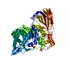 4ekjS S: Starting model for refinement |
|---|---|
| Similar structure data |
-Links
 PDBj
PDBj- Assembly
| Deposited unit | 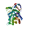
| ||||||||
|---|---|---|---|---|---|---|---|---|---|
| 1 |
| ||||||||
| Unit cell |
|
-Components
| #1: Protein | Xylan 1,4-b-xylosidase Mass: 56305.574 Da / Num. of mol.: 1 Source method: isolated from a genetically manipulated source Source: (gene. exp.) Caulobacter crescentus (bacteria) / Strain: ATCC 19089 / CB15 / Gene: Itgav, CC_2357 / Production host: Escherichia coli (E. coli) / References: UniProt: Q9A5U0 |
|---|---|
| #2: Chemical | ChemComp-MES / MES (buffer)  Mass: 195.237 Da / Num. of mol.: 1 / Source method: obtained synthetically / Formula: C6H13NO4S / Comment: pH buffer*YM Mass: 195.237 Da / Num. of mol.: 1 / Source method: obtained synthetically / Formula: C6H13NO4S / Comment: pH buffer*YM |
| #3: Water | ChemComp-HOH / Water Mass: 18.015 Da / Num. of mol.: 149 / Source method: isolated from a natural source / Formula: H2O Mass: 18.015 Da / Num. of mol.: 149 / Source method: isolated from a natural source / Formula: H2O |
-Experimental details
-Experiment
| Experiment | Method: X-RAY DIFFRACTION / Number of used crystals: 1 |
|---|
- Sample preparation
| Crystal | Density Matthews: 2.02 Å3/Da / Density % sol: 39.25 % |
|---|---|
| Crystal grow | Temperature: 293 K / Method: vapor diffusion, hanging drop / pH: 6 Details: 0.1 M MES, 12% PEG6000, 0.05 M NaCl, pH 6.0, VAPOR DIFFUSION, HANGING DROP, temperature 293K |
-Data collection
| Diffraction | Mean temperature: 100 K |
|---|---|
| Diffraction source | Source: SYNCHROTRON / Site: LNLS  / Beamline: W01B-MX2 / Wavelength: 1.6 Å / Beamline: W01B-MX2 / Wavelength: 1.6 Å |
| Detector | Type: MARMOSAIC 225 mm CCD / Detector: CCD / Date: Mar 5, 2012 |
| Radiation | Monochromator: Si111 / Protocol: SINGLE WAVELENGTH / Monochromatic (M) / Laue (L): M / Scattering type: x-ray |
| Radiation wavelength | Wavelength: 1.6 Å / Relative weight: 1 |
| Reflection | Resolution: 2.1→17.4 Å / Num. all: 25413 / Num. obs: 25082 / % possible obs: 96.2 % / Observed criterion σ(F): 0 / Observed criterion σ(I): 1 |
| Reflection shell | Resolution: 2.1→2.14 Å / % possible all: 96.8 |
- Processing
| Software |
| ||||||||||||||||||||||||||||||||||||||||||||||||||||||||||||
|---|---|---|---|---|---|---|---|---|---|---|---|---|---|---|---|---|---|---|---|---|---|---|---|---|---|---|---|---|---|---|---|---|---|---|---|---|---|---|---|---|---|---|---|---|---|---|---|---|---|---|---|---|---|---|---|---|---|---|---|---|---|
| Refinement | Method to determine structure: MOLECULAR REPLACEMENT Starting model: PDB entry 4EKJ Resolution: 2.1→17.4 Å / Cor.coef. Fo:Fc: 0.895 / Cor.coef. Fo:Fc free: 0.846 / SU B: 11.983 / SU ML: 0.163 / Cross valid method: THROUGHOUT / ESU R: 0.313 / ESU R Free: 0.243 / Stereochemistry target values: MAXIMUM LIKELIHOOD
| ||||||||||||||||||||||||||||||||||||||||||||||||||||||||||||
| Solvent computation | Ion probe radii: 0.8 Å / Shrinkage radii: 0.8 Å / VDW probe radii: 1.2 Å / Solvent model: MASK | ||||||||||||||||||||||||||||||||||||||||||||||||||||||||||||
| Displacement parameters | Biso mean: 16.78 Å2
| ||||||||||||||||||||||||||||||||||||||||||||||||||||||||||||
| Refinement step | Cycle: LAST / Resolution: 2.1→17.4 Å
| ||||||||||||||||||||||||||||||||||||||||||||||||||||||||||||
| Refine LS restraints |
| ||||||||||||||||||||||||||||||||||||||||||||||||||||||||||||
| LS refinement shell | Resolution: 2.1→2.155 Å / Total num. of bins used: 20
| ||||||||||||||||||||||||||||||||||||||||||||||||||||||||||||
| Refinement TLS params. | Method: refined / Origin x: -10.2259 Å / Origin y: -3.0899 Å / Origin z: -23.8065 Å
|
