 Movie
Movie Controller


+ Open data

- Basic information
| Entry | Database: PDB / ID: 4m0d | ||||||
|---|---|---|---|---|---|---|---|
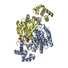
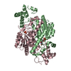
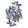
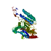
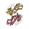
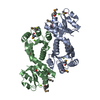
| Title | Crystal structure of MurQ from H.influenzae in apo form | ||||||
 Components Components | N-acetylmuramic acid 6-phosphate etherase | ||||||
 Keywords Keywords | LYASE / NAD(P)/FAD- binding Rossmann fold / Alpha-Beta-Alpha sandwich / MurQ / YfeU | ||||||
| Function / homology |  Function and homology informationN-acetylmuramic acid 6-phosphate etherase / N-acetylmuramic acid catabolic process / 1,6-anhydro-N-acetyl-beta-muramic acid catabolic process / amino sugar catabolic process / ether hydrolase activity / carbon-oxygen lyase activity / fructose-6-phosphate binding / peptidoglycan turnover / response to fructose / enzyme inhibitor activity ...N-acetylmuramic acid 6-phosphate etherase / N-acetylmuramic acid catabolic process / 1,6-anhydro-N-acetyl-beta-muramic acid catabolic process / amino sugar catabolic process / ether hydrolase activity / carbon-oxygen lyase activity / fructose-6-phosphate binding / peptidoglycan turnover / response to fructose / enzyme inhibitor activity / glucose homeostasis / carbohydrate binding / enzyme binding / cytosol Function and homology informationN-acetylmuramic acid 6-phosphate etherase / N-acetylmuramic acid catabolic process / 1,6-anhydro-N-acetyl-beta-muramic acid catabolic process / amino sugar catabolic process / ether hydrolase activity / carbon-oxygen lyase activity / fructose-6-phosphate binding / peptidoglycan turnover / response to fructose / enzyme inhibitor activity ...N-acetylmuramic acid 6-phosphate etherase / N-acetylmuramic acid catabolic process / 1,6-anhydro-N-acetyl-beta-muramic acid catabolic process / amino sugar catabolic process / ether hydrolase activity / carbon-oxygen lyase activity / fructose-6-phosphate binding / peptidoglycan turnover / response to fructose / enzyme inhibitor activity / glucose homeostasis / carbohydrate binding / enzyme binding / cytosolSimilarity search - Function | ||||||
| Biological species |  Haemophilus influenzae (bacteria) Haemophilus influenzae (bacteria) | ||||||
| Method | X-RAY DIFFRACTION / MOLECULAR REPLACEMENT / Resolution: 2.577 Å | ||||||
 Authors Authors | Hazra, S. / Blanchard, J. | ||||||
 Citation Citation | Journal: Biochemistry / Year: 2013 Title: Structure of MurNAc 6-phosphate hydrolase (MurQ) from Haemophilus influenzae with a bound inhibitor. Authors: Hadi, T. / Hazra, S. / Tanner, M.E. / Blanchard, J.S. | ||||||
| History |
|
- Structure visualization
| Structure viewer | Molecule: MolmilJmol/JSmol |
|---|
- Downloads & links
-Download
| PDBx/mmCIF format | 4m0d.cif.gz | 227.9 KB | Display | PDBx/mmCIF format |
|---|---|---|---|---|
| PDB format | pdb4m0d.ent.gz | 185.3 KB | Display | PDB format |
| PDBx/mmJSON format | 4m0d.json.gz | Tree view | PDBx/mmJSON format | |
| Others |  Other downloads Other downloads |
-Validation report
| Arichive directory | https://data.pdbj.org/pub/pdb/validation_reports/m0/4m0dftp://data.pdbj.org/pub/pdb/validation_reports/m0/4m0d | HTTPS FTP |
|---|
-Related structure data
| Related structure data |  4lzjC  1nriS C: citing same article ( S: Starting model for refinement |
|---|---|
| Similar structure data |


-Links
 PDBj
PDBj- Assembly
| Deposited unit | 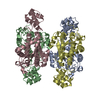
| ||||||||
|---|---|---|---|---|---|---|---|---|---|
| 1 | 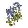
| ||||||||
| 2 | 
| ||||||||
| 3 |
| ||||||||
| Unit cell |
|
-Components
| #1: Protein | / MurNAc-6-P etherase / N-acetylmuramic acid 6-phosphate hydrolase / N-acetylmuramic acid 6-phosphate lyase Mass: 32564.734 Da / Num. of mol.: 4 Source method: isolated from a genetically manipulated source Source: (gene. exp.) Haemophilus influenzae (bacteria) / Strain: Rd KW20 / Gene: murQ / Production host: Escherichia coli (E. coli)References: UniProt: P44862, N-acetylmuramic acid 6-phosphate etherase#2: Water | ChemComp-HOH / | Water Mass: 18.015 Da / Num. of mol.: 283 / Source method: isolated from a natural source / Formula: H2O Mass: 18.015 Da / Num. of mol.: 283 / Source method: isolated from a natural source / Formula: H2O |
|---|
-Experimental details
-Experiment
| Experiment | Method: X-RAY DIFFRACTION / Number of used crystals: 1 |
|---|
- Sample preparation
| Crystal | Density Matthews: 2.19 Å3/Da / Density % sol: 43.94 % |
|---|---|
| Crystal grow | Temperature: 298 K / Method: vapor diffusion, sitting drop / pH: 5.5 Details: 0.2 M Nacl, 0.1 M Bis-Tris:HCl, pH 5.5, 25% (w/v) PEG-3350, VAPOR DIFFUSION, SITTING DROP, temperature 298.0K |
-Data collection
| Diffraction | Mean temperature: 65 K |
|---|---|
| Diffraction source | Source: ROTATING ANODE / Type: RIGAKU / Wavelength: 1.5418 Å |
| Detector | Detector: IMAGE PLATE |
| Radiation | Monochromator: GRAPHITE / Protocol: SINGLE WAVELENGTH / Monochromatic (M) / Laue (L): M / Scattering type: x-ray |
| Radiation wavelength | Wavelength: 1.5418 Å / Relative weight: 1 |
| Reflection | Resolution: 2.58→32.56 Å / Num. all: 35231 / Num. obs: 34265 / Redundancy: 5.5 % |
| Reflection shell | Highest resolution: 2.58 Å |
- Processing
| Software |
| |||||||||||||||||||||||||||||||||||||||||||||||||||||||||||||||||||||||||||||||||||||||||||||||||||||||||
|---|---|---|---|---|---|---|---|---|---|---|---|---|---|---|---|---|---|---|---|---|---|---|---|---|---|---|---|---|---|---|---|---|---|---|---|---|---|---|---|---|---|---|---|---|---|---|---|---|---|---|---|---|---|---|---|---|---|---|---|---|---|---|---|---|---|---|---|---|---|---|---|---|---|---|---|---|---|---|---|---|---|---|---|---|---|---|---|---|---|---|---|---|---|---|---|---|---|---|---|---|---|---|---|---|---|---|
| Refinement | Method to determine structure: MOLECULAR REPLACEMENT Starting model: 1NRI Resolution: 2.577→32.563 Å / SU ML: 0.42 / σ(F): 0.11 / Phase error: 30.38 / Stereochemistry target values: ML
| |||||||||||||||||||||||||||||||||||||||||||||||||||||||||||||||||||||||||||||||||||||||||||||||||||||||||
| Solvent computation | Shrinkage radii: 0.9 Å / VDW probe radii: 1.11 Å / Solvent model: FLAT BULK SOLVENT MODEL | |||||||||||||||||||||||||||||||||||||||||||||||||||||||||||||||||||||||||||||||||||||||||||||||||||||||||
| Refinement step | Cycle: LAST / Resolution: 2.577→32.563 Å
| |||||||||||||||||||||||||||||||||||||||||||||||||||||||||||||||||||||||||||||||||||||||||||||||||||||||||
| Refine LS restraints |
| |||||||||||||||||||||||||||||||||||||||||||||||||||||||||||||||||||||||||||||||||||||||||||||||||||||||||
| LS refinement shell |
|
