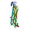
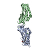
A: Short palate, lung and nasal epithelium carcinoma-associated protein 2B B: Short palate, lung and nasal epithelium carcinoma-associated protein 2B hetero molecules
Resolution: 2.3→48.7 Å / Cor.coef. Fo:Fc: 0.948 / Cor.coef. Fo:Fc free: 0.935 / SU B: 20.376 / SU ML: 0.228 / Cross valid method: THROUGHOUT / ESU R: 0.355 / ESU R Free: 0.25 Details: U VALUES : WITH TLS ADDED HYDROGENS HAVE BEEN ADDED IN THE RIDING POSITIONS
Rfactor
Num. reflection
% reflection
Selection details
Rfree
0.26505
980
5 %
RANDOM
Rwork
0.22311
-
-
-
obs
0.22525
18652
99.32 %
-
Solvent computation
Ion probe radii: 0.7 Å / Shrinkage radii: 0.7 Å / VDW probe radii: 1 Å
Displacement parameters
Biso mean: 69.715 Å2
Baniso -1
Baniso -2
Baniso -3
1-
-2.7 Å2
-0 Å2
-0.95 Å2
2-
-
5.16 Å2
-0 Å2
3-
-
-
-1.54 Å2
Refinement step
Cycle: LAST / Resolution: 2.3→48.7 Å
Protein
Nucleic acid
Ligand
Solvent
Total
Num. atoms
2930
0
43
51
3024
Refine LS restraints
Refine-ID
Type
Dev ideal
Dev ideal target
Number
X-RAY DIFFRACTION
r_bond_refined_d
0.007
0.013
2990
X-RAY DIFFRACTION
r_bond_other_d
0.003
0.017
2926
X-RAY DIFFRACTION
r_angle_refined_deg
1.407
1.636
4045
X-RAY DIFFRACTION
r_angle_other_deg
1.219
1.567
6790
X-RAY DIFFRACTION
r_dihedral_angle_1_deg
6.876
5
373
X-RAY DIFFRACTION
r_dihedral_angle_2_deg
34.649
23.125
160
X-RAY DIFFRACTION
r_dihedral_angle_3_deg
16.171
15
549
X-RAY DIFFRACTION
r_dihedral_angle_4_deg
17.178
15
23
X-RAY DIFFRACTION
r_chiral_restr
0.064
0.2
416
X-RAY DIFFRACTION
r_gen_planes_refined
0.006
0.02
3268
X-RAY DIFFRACTION
r_gen_planes_other
0.001
0.02
545
X-RAY DIFFRACTION
r_mcbond_it
1.037
2.792
1507
X-RAY DIFFRACTION
r_mcbond_other
1.03
2.791
1506
X-RAY DIFFRACTION
r_mcangle_it
1.73
4.176
1875
X-RAY DIFFRACTION
r_mcangle_other
1.729
4.178
1876
X-RAY DIFFRACTION
r_scbond_it
1.302
3.151
1483
X-RAY DIFFRACTION
r_scbond_other
1.302
3.153
1484
X-RAY DIFFRACTION
r_scangle_other
2.119
4.59
2171
X-RAY DIFFRACTION
r_long_range_B_refined
7.108
34.58
3093
X-RAY DIFFRACTION
r_long_range_B_other
7.105
34.493
3087
LS refinement shell
Resolution: 2.3→2.36 Å / Total num. of bins used: 20
Rfactor
Num. reflection
% reflection
Rfree
0.365
82
-
Rwork
0.276
1400
-
obs
-
-
99.26 %
Refinement TLS params.
Method: refined / Refine-ID: X-RAY DIFFRACTION
ID
L11 (°2)
L12 (°2)
L13 (°2)
L22 (°2)
L23 (°2)
L33 (°2)
S11 (Å °)
S12 (Å °)
S13 (Å °)
S21 (Å °)
S22 (Å °)
S23 (Å °)
S31 (Å °)
S32 (Å °)
S33 (Å °)
T11 (Å2)
T12 (Å2)
T13 (Å2)
T22 (Å2)
T23 (Å2)
T33 (Å2)
Origin x (Å)
Origin y (Å)
Origin z (Å)
1
3.6016
-0.0108
2.092
0.9866
-0.7943
7.182
0.1246
0.0686
0.1363
0.2449
-0.0602
-0.1895
0.6544
0.6806
-0.0644
0.6862
0.1151
-0.0983
0.0902
-0.0446
0.0963
78.257
39.418
31.284
2
6.7654
0.5888
2.7897
1.4957
0.091
3.9078
0.3184
-0.399
0.0119
0.1558
-0.2914
0.1769
0.3463
-0.5876
-0.027
0.5626
-0.0073
-0.0626
0.2184
-0.113
0.1131
72.379
19.775
12.359
Refinement TLS group
ID
Refine-ID
Refine TLS-ID
Auth asym-ID
Auth seq-ID
1
X-RAY DIFFRACTION
1
A
38 - 238
2
X-RAY DIFFRACTION
2
B
38 - 237
+
About Yorodumi
-
News
-
Feb 9, 2022. New format data for meta-information of EMDB entries
New format data for meta-information of EMDB entries
Version 3 of the EMDB header file is now the official format.
The previous official version 1.9 will be removed from the archive.
In the structure databanks used in Yorodumi, some data are registered as the other names, "COVID-19 virus" and "2019-nCoV". Here are the details of the virus and the list of structure data.
Jan 31, 2019. EMDB accession codes are about to change! (news from PDBe EMDB page)
EMDB accession codes are about to change! (news from PDBe EMDB page)
The allocation of 4 digits for EMDB accession codes will soon come to an end. Whilst these codes will remain in use, new EMDB accession codes will include an additional digit and will expand incrementally as the available range of codes is exhausted. The current 4-digit format prefixed with “EMD-” (i.e. EMD-XXXX) will advance to a 5-digit format (i.e. EMD-XXXXX), and so on. It is currently estimated that the 4-digit codes will be depleted around Spring 2019, at which point the 5-digit format will come into force.
The EM Navigator/Yorodumi systems omit the EMD- prefix.
Related info.:Q: What is EMD? / ID/Accession-code notation in Yorodumi/EM Navigator
Yorodumi is a browser for structure data from EMDB, PDB, SASBDB, etc.
This page is also the successor to EM Navigator detail page, and also detail information page/front-end page for Omokage search.
The word "yorodu" (or yorozu) is an old Japanese word meaning "ten thousand". "mi" (miru) is to see.
Related info.:EMDB / PDB / SASBDB / Comparison of 3 databanks / Yorodumi Search / Aug 31, 2016. New EM Navigator & Yorodumi / Yorodumi Papers / Jmol/JSmol / Function and homology information / Changes in new EM Navigator and Yorodumi
 Movie
Movie Controller


 Open data
 Basic information
 Components
 Keywords
 Lipid-binding serum glycoprotein, N-terminal / Bactericidal permeability-increasing protein, alpha/beta domain superfamily / LBP / BPI / CETP family, N-terminal domain /
  Function and homology information
 Authors
 Citation
 Structure visualization
 Downloads & links
 Other downloads

 PDBj
PDBj


 Assembly


 Mass: 40.078 Da / Num. of mol.: 3 / Source method: obtained synthetically / Formula: Ca
 Mass: 282.461 Da / Num. of mol.: 2 / Source method: obtained synthetically / Formula: C18H34O2 / Feature type: SUBJECT OF INVESTIGATION
 Mass: 18.015 Da / Num. of mol.: 51 / Source method: isolated from a natural source / Formula: H2O
 Sample preparation
 / Beamline: MX2 / Wavelength: 0.9537 Å
 Processing