 Movie
Movie Controller

[English] 日本語
 Yorodumi
- PDB-4lvu: Crystal Structure of a Putative Short Chain Dehydrogenase from Bu... -
+ Open data

- Basic information
| Entry | Database: PDB / ID: 4lvu | ||||||
|---|---|---|---|---|---|---|---|
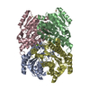
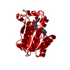
| Title | Crystal Structure of a Putative Short Chain Dehydrogenase from Burkholderia thailandensis | ||||||
 Components Components | Oxidoreductase, short chain dehydrogenase/reductase family | ||||||
 Keywords Keywords | OXIDOREDUCTASE / Structural Genomics / NIAID / National Institute of Allergy and Infectious Diseases / Seattle Structural Genomics Center for Infectious Disease / SSGCID | ||||||
| Function / homology |  Function and homology information Function and homology information | ||||||
| Biological species |  Burkholderia thailandensis (bacteria) Burkholderia thailandensis (bacteria) | ||||||
| Method | X-RAY DIFFRACTION / SYNCHROTRON / MOLECULAR REPLACEMENT / molecular replacement / Resolution: 1.25 Å | ||||||
 Authors Authors | Seattle Structural Genomics Center for Infectious Disease (SSGCID) | ||||||
 Citation Citation | Journal: TO BE PUBLISHED Title: Crystal Structure of a Putative Short Chain Dehydrogenase from Burkholderia thailandensis Authors: Dranow, D.M. / Edwards, T.E. / Lorimer, D. | ||||||
| History |
|
- Structure visualization
| Structure viewer | Molecule: MolmilJmol/JSmol |
|---|
- Downloads & links
-Download
| PDBx/mmCIF format | 4lvu.cif.gz | 227.2 KB | Display | PDBx/mmCIF format |
|---|---|---|---|---|
| PDB format | pdb4lvu.ent.gz | 182.1 KB | Display | PDB format |
| PDBx/mmJSON format | 4lvu.json.gz | Tree view | PDBx/mmJSON format | |
| Others |  Other downloads Other downloads |
-Validation report
| Arichive directory | https://data.pdbj.org/pub/pdb/validation_reports/lv/4lvuftp://data.pdbj.org/pub/pdb/validation_reports/lv/4lvu | HTTPS FTP |
|---|
-Related structure data
| Related structure data |  4jroS S: Starting model for refinement |
|---|---|
| Similar structure data | |
| Other databases |


-Links
 PDBj
PDBj


- Assembly
| Deposited unit | 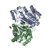
| ||||||||
|---|---|---|---|---|---|---|---|---|---|
| 1 | 
| ||||||||
| Unit cell |
|
-Components
| #1: Protein | Mass: 29740.822 Da / Num. of mol.: 2 Source method: isolated from a genetically manipulated source Source: (gene. exp.) Burkholderia thailandensis (bacteria) / Strain: E264 / ATCC 700388 / DSM 13276 / CIP 106301 / Gene: BTH_I1182 / Production host: Escherichia coli (E. coli) / References: UniProt: Q2SZC0, Oxidoreductases#2: Chemical | Ethylene glycol  Mass: 62.068 Da / Num. of mol.: 2 / Source method: obtained synthetically / Formula: C2H6O2 Mass: 62.068 Da / Num. of mol.: 2 / Source method: obtained synthetically / Formula: C2H6O2#3: Water | ChemComp-HOH / | Water Mass: 18.015 Da / Num. of mol.: 671 / Source method: isolated from a natural source / Formula: H2O Mass: 18.015 Da / Num. of mol.: 671 / Source method: isolated from a natural source / Formula: H2O |
|---|
-Experimental details
-Experiment
| Experiment | Method: X-RAY DIFFRACTION / Number of used crystals: 1 |
|---|
- Sample preparation
| Crystal | Density Matthews: 2.21 Å3/Da / Density % sol: 44.44 % |
|---|---|
| Crystal grow | Temperature: 289 K / Method: vapor diffusion, sitting drop Details: JCSG+(a5): 20% PEG3350, 200 mM magnesium formate, VAPOR DIFFUSION, SITTING DROP, temperature 289K |
-Data collection
| Diffraction | Mean temperature: 100 K | |||||||||||||||||||||||||||||||||||||||||||||||||||||||||||||||||||||||||||||||||||||||||||||||||||||||||||||||||||||||||||||||||||||||||||||||||||
|---|---|---|---|---|---|---|---|---|---|---|---|---|---|---|---|---|---|---|---|---|---|---|---|---|---|---|---|---|---|---|---|---|---|---|---|---|---|---|---|---|---|---|---|---|---|---|---|---|---|---|---|---|---|---|---|---|---|---|---|---|---|---|---|---|---|---|---|---|---|---|---|---|---|---|---|---|---|---|---|---|---|---|---|---|---|---|---|---|---|---|---|---|---|---|---|---|---|---|---|---|---|---|---|---|---|---|---|---|---|---|---|---|---|---|---|---|---|---|---|---|---|---|---|---|---|---|---|---|---|---|---|---|---|---|---|---|---|---|---|---|---|---|---|---|---|---|---|---|
| Diffraction source | Source: SYNCHROTRON / Site: APS  / Beamline: 21-ID-F / Wavelength: 0.9786 Å / Beamline: 21-ID-F / Wavelength: 0.9786 Å | |||||||||||||||||||||||||||||||||||||||||||||||||||||||||||||||||||||||||||||||||||||||||||||||||||||||||||||||||||||||||||||||||||||||||||||||||||
| Detector | Type: MARMOSAIC 225 mm CCD / Detector: CCD / Date: Jun 19, 2013 / Details: beryllium lenses | |||||||||||||||||||||||||||||||||||||||||||||||||||||||||||||||||||||||||||||||||||||||||||||||||||||||||||||||||||||||||||||||||||||||||||||||||||
| Radiation | Monochromator: diamond(111) / Protocol: SINGLE WAVELENGTH / Monochromatic (M) / Laue (L): M / Scattering type: x-ray | |||||||||||||||||||||||||||||||||||||||||||||||||||||||||||||||||||||||||||||||||||||||||||||||||||||||||||||||||||||||||||||||||||||||||||||||||||
| Radiation wavelength | Wavelength: 0.9786 Å / Relative weight: 1 | |||||||||||||||||||||||||||||||||||||||||||||||||||||||||||||||||||||||||||||||||||||||||||||||||||||||||||||||||||||||||||||||||||||||||||||||||||
| Reflection | Resolution: 1.25→50 Å / Num. all: 146259 / Num. obs: 143334 / % possible obs: 98 % / Observed criterion σ(F): 0 / Observed criterion σ(I): -3 / Redundancy: 9.8 % / Biso Wilson estimate: 14.702 Å2 / Rmerge(I) obs: 0.05 / Net I/σ(I): 26.37 | |||||||||||||||||||||||||||||||||||||||||||||||||||||||||||||||||||||||||||||||||||||||||||||||||||||||||||||||||||||||||||||||||||||||||||||||||||
| Reflection shell | Diffraction-ID: 1
|
-Phasing
| Phasing | Method: molecular replacement | |||||||||
|---|---|---|---|---|---|---|---|---|---|---|
| Phasing MR | Model details: Phaser MODE: MR_AUTO
|
- Processing
| Software |
| ||||||||||||||||||||||||||||||||||||||||||||||||||||||||||||||||||||||||||||||||||||||||||
|---|---|---|---|---|---|---|---|---|---|---|---|---|---|---|---|---|---|---|---|---|---|---|---|---|---|---|---|---|---|---|---|---|---|---|---|---|---|---|---|---|---|---|---|---|---|---|---|---|---|---|---|---|---|---|---|---|---|---|---|---|---|---|---|---|---|---|---|---|---|---|---|---|---|---|---|---|---|---|---|---|---|---|---|---|---|---|---|---|---|---|---|
| Refinement | Method to determine structure: MOLECULAR REPLACEMENT Starting model: PDB ENTRY 4JRO Resolution: 1.25→45.4 Å / Cor.coef. Fo:Fc: 0.979 / Cor.coef. Fo:Fc free: 0.971 / WRfactor Rfree: 0.1361 / WRfactor Rwork: 0.1137 / Occupancy max: 1 / Occupancy min: 0.3 / FOM work R set: 0.9382 / SU B: 0.922 / SU ML: 0.019 / SU R Cruickshank DPI: 0.0325 / SU Rfree: 0.0329 / Cross valid method: THROUGHOUT / σ(F): 0 / ESU R: 0.033 / ESU R Free: 0.033 / Stereochemistry target values: MAXIMUM LIKELIHOOD / Details: HYDROGENS HAVE BEEN ADDED IN THE RIDING POSITIONS
| ||||||||||||||||||||||||||||||||||||||||||||||||||||||||||||||||||||||||||||||||||||||||||
| Solvent computation | Ion probe radii: 0.8 Å / Shrinkage radii: 0.8 Å / VDW probe radii: 1.2 Å / Solvent model: MASK | ||||||||||||||||||||||||||||||||||||||||||||||||||||||||||||||||||||||||||||||||||||||||||
| Displacement parameters | Biso max: 115.42 Å2 / Biso mean: 13.3299 Å2 / Biso min: 3.97 Å2
| ||||||||||||||||||||||||||||||||||||||||||||||||||||||||||||||||||||||||||||||||||||||||||
| Refinement step | Cycle: LAST / Resolution: 1.25→45.4 Å
| ||||||||||||||||||||||||||||||||||||||||||||||||||||||||||||||||||||||||||||||||||||||||||
| Refine LS restraints |
| ||||||||||||||||||||||||||||||||||||||||||||||||||||||||||||||||||||||||||||||||||||||||||
| LS refinement shell | Resolution: 1.25→1.282 Å / Total num. of bins used: 20
|
