Mass: 18.015 Da / Num. of mol.: 633 / Source method: isolated from a natural source / Formula: H2O
-
Details
Sequence details
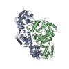
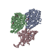
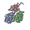
THE SEQUENCE OF KINESIN (CHAIN K) INCLUDED THE FOLLOWING MUTATIONS: C7S, C65A, C168A, C174S AND ...THE SEQUENCE OF KINESIN (CHAIN K) INCLUDED THE FOLLOWING MUTATIONS: C7S, C65A, C168A, C174S AND C294A. THE CONSTRUCT IS KNOWN AS CYS-LITE (RICE ET AL, NATURE 1999, VOL 402: 778-84). IT WAS TRUNCATED AFTER RESIDUE 325 FOR THIS STUDY. FOR TUBULIN THE BOVINE BRAIN TUBULIN SEQUENCE WAS USED FOR REFINEMENT BECAUSE THE SEQUENCE OF OVINE BRAIN TUBULIN IS NOT AVAILABLE. FOR ALPHA-TUBULIN (CHAIN A) THE ALPHA 1B ISOTYPE SEQUENCE (NCBI NP_001108328.1) WAS USED. FOR BETA-TUBULIN (CHAIN B), THERE ARE THREE MAJOR ISOTYPES EXPRESSED IN THE BRAIN (BANERJEE AT AL, JBC 1988, VOL 263:3029-34). THE BETA 2B ISOTYPE SEQUENCE (NCBI NP_001003900.1) WAS USED BUT POINT MUTATIONS WERE INTRODUCED WHEN POSSIBLE TO TAKE INTO ACCOUNT THE ISOTYPE DIVERSITY.
-
Experimental details
-
Experiment
Experiment
Method: X-RAY DIFFRACTION / Number of used crystals: 1
-
Sample preparation
Crystal
Density Matthews: 2.74 Å3/Da / Density % sol: 55 %
Crystal grow
Temperature: 293 K / Method: vapor diffusion / pH: 6.5 Details: PEG, MES BUFFER, 0.2M AMMONIUM SULFATE, pH 6.50, VAPOR DIFFUSION, temperature 293K
In the structure databanks used in Yorodumi, some data are registered as the other names, "COVID-19 virus" and "2019-nCoV". Here are the details of the virus and the list of structure data.
Jan 31, 2019. EMDB accession codes are about to change! (news from PDBe EMDB page)
EMDB accession codes are about to change! (news from PDBe EMDB page)
The allocation of 4 digits for EMDB accession codes will soon come to an end. Whilst these codes will remain in use, new EMDB accession codes will include an additional digit and will expand incrementally as the available range of codes is exhausted. The current 4-digit format prefixed with “EMD-” (i.e. EMD-XXXX) will advance to a 5-digit format (i.e. EMD-XXXXX), and so on. It is currently estimated that the 4-digit codes will be depleted around Spring 2019, at which point the 5-digit format will come into force.
The EM Navigator/Yorodumi systems omit the EMD- prefix.
Related info.:Q: What is EMD? / ID/Accession-code notation in Yorodumi/EM Navigator
Yorodumi is a browser for structure data from EMDB, PDB, SASBDB, etc.
This page is also the successor to EM Navigator detail page, and also detail information page/front-end page for Omokage search.
The word "yorodu" (or yorozu) is an old Japanese word meaning "ten thousand". "mi" (miru) is to see.
Related info.:EMDB / PDB / SASBDB / Comparison of 3 databanks / Yorodumi Search / Aug 31, 2016. New EM Navigator & Yorodumi / Yorodumi Papers / Jmol/JSmol / Function and homology information / Changes in new EM Navigator and Yorodumi
 Movie
Movie Controller

 Yorodumi
 Open data
 Basic information
 Components
 Keywords
 BETA-TUBULIN /
  Function and homology information

 Authors
 Citation
 Structure visualization
 Downloads & links
 Other downloads
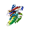


 PDBj
PDBj


 Assembly
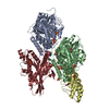


 Mass: 523.180 Da / Num. of mol.: 1 / Source method: obtained synthetically / Formula: C10H16N5O14P3 / Comment: GTP, energy-carrying molecule*YM
 Mass: 24.305 Da / Num. of mol.: 1 / Source method: obtained synthetically / Formula: Mg
 Mass: 96.063 Da / Num. of mol.: 11 / Source method: obtained synthetically / Formula: SO4
 Type: RNA linking / Mass: 443.201 Da / Num. of mol.: 1 / Source method: obtained synthetically / Formula: C10H15N5O11P2 / Comment: GDP, energy-carrying molecule*YM
 Mass: 195.237 Da / Num. of mol.: 1 / Source method: obtained synthetically / Formula: C6H13NO4S / Comment: pH buffer*YM
 Mass: 92.094 Da / Num. of mol.: 4 / Source method: obtained synthetically / Formula: C3H8O3
 Sample preparation
 / Beamline: PROXIMA 1 / Wavelength: 0.979 / Wavelength: 0.979 Å
 Processing