 Movie
Movie Controller

[English] 日本語
 Yorodumi
- PDB-4leq: tRNA guanine transglycosylase (TGT) in complex with Furanoside-Ba... -
+ Open data

- Basic information
| Entry | Database: PDB / ID: 4leq | ||||||
|---|---|---|---|---|---|---|---|
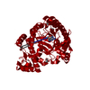
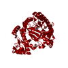
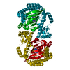
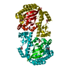
| Title | tRNA guanine transglycosylase (TGT) in complex with Furanoside-Based lin-Benzoguanine 1 | ||||||
 Components Components | Queuine tRNA-ribosyltransferase | ||||||
 Keywords Keywords | TRANSFERASE/TRANSFERASE INHIBITOR / Tim Barrel / GLYCOSYLTRANSFERASE / Zinc-BINDING / TRANSFERASE / TRANSFERASE-TRANSFERASE INHIBITOR complex | ||||||
| Function / homology |  Function and homology information Function and homology informationtRNA-guanosine34 preQ1 transglycosylase / tRNA-guanosine(34) queuine transglycosylase activity / tRNA-guanine transglycosylation / queuosine biosynthetic process / metal ion bindingSimilarity search - Function | ||||||
| Biological species |  Zymomonas Mobilis subsp. mobilis (bacteria) Zymomonas Mobilis subsp. mobilis (bacteria) | ||||||
| Method | X-RAY DIFFRACTION / SYNCHROTRON / MOLECULAR REPLACEMENT / Resolution: 1.405 Å | ||||||
 Authors Authors | Ehrmann, F.R. / Heine, A. / Klebe, G. | ||||||
 Citation Citation | Journal: Chemistry / Year: 2015 Title: Replacement of Water Molecules in a Phosphate Binding Site by Furanoside-Appended lin-Benzoguanine Ligands of tRNA-Guanine Transglycosylase (TGT). Authors: Barandun, L.J. / Ehrmann, F.R. / Zimmerli, D. / Immekus, F. / Giroud, M. / Grunenfelder, C. / Schweizer, W.B. / Bernet, B. / Betz, M. / Heine, A. / Klebe, G. / Diederich, F. | ||||||
| History |
|
- Structure visualization
| Structure viewer | Molecule: MolmilJmol/JSmol |
|---|
- Downloads & links
-Download
| PDBx/mmCIF format | 4leq.cif.gz | 181.7 KB | Display | PDBx/mmCIF format |
|---|---|---|---|---|
| PDB format | pdb4leq.ent.gz | 143.5 KB | Display | PDB format |
| PDBx/mmJSON format | 4leq.json.gz | Tree view | PDBx/mmJSON format | |
| Others |  Other downloads Other downloads |
-Validation report
| Arichive directory | https://data.pdbj.org/pub/pdb/validation_reports/le/4leqftp://data.pdbj.org/pub/pdb/validation_reports/le/4leq | HTTPS FTP |
|---|
-Related structure data
| Related structure data |  4kwoC  4lbuC  1pudS C: citing same article ( S: Starting model for refinement |
|---|---|
| Similar structure data |


-Links
 PDBj
PDBj- Assembly
| Deposited unit | 
| |||||||||
|---|---|---|---|---|---|---|---|---|---|---|
| 1 | 
| |||||||||
| Unit cell |
| |||||||||
| Components on special symmetry positions |
|
-Components
-Protein , 1 types, 1 molecules A
| #1: Protein | / Guanine insertion enzyme / tRNA-guanine transglycosylase Mass: 43069.828 Da / Num. of mol.: 1 Source method: isolated from a genetically manipulated source Source: (gene. exp.) Zymomonas Mobilis subsp. mobilis (bacteria)Strain: ATCC 31821 / ZM4 / CP4 / Gene: tgt, ZMO0363 / Plasmid: pPR-IBA2 / Production host: Escherichia coli (E. coli) / Strain (production host): BL21(DE3)CodonPlusReferences: UniProt: P28720, tRNA-guanosine34 preQ1 transglycosylase |
|---|
-Non-polymers , 5 types, 433 molecules 


| #2: Chemical | ChemComp-1WK /  Mass: 390.394 Da / Num. of mol.: 1 / Source method: obtained synthetically / Formula: C17H22N6O5 Mass: 390.394 Da / Num. of mol.: 1 / Source method: obtained synthetically / Formula: C17H22N6O5 | ||||||
|---|---|---|---|---|---|---|---|
| #3: Chemical | Glycerol Mass: 92.094 Da / Num. of mol.: 2 / Source method: obtained synthetically / Formula: C3H8O3 Mass: 92.094 Da / Num. of mol.: 2 / Source method: obtained synthetically / Formula: C3H8O3#4: Chemical | ChemComp-ZN / |  Mass: 65.409 Da / Num. of mol.: 1 / Source method: obtained synthetically / Formula: Zn Mass: 65.409 Da / Num. of mol.: 1 / Source method: obtained synthetically / Formula: Zn#5: Chemical | ChemComp-CL / | Chloride Mass: 35.453 Da / Num. of mol.: 1 / Source method: obtained synthetically / Formula: Cl Mass: 35.453 Da / Num. of mol.: 1 / Source method: obtained synthetically / Formula: Cl#6: Water | ChemComp-HOH / | WaterMass: 18.015 Da / Num. of mol.: 428 / Source method: isolated from a natural source / Formula: H2O |
-Experimental details
-Experiment
| Experiment | Method: X-RAY DIFFRACTION / Number of used crystals: 1 |
|---|
- Sample preparation
| Crystal | Density Matthews: 2.39 Å3/Da / Density % sol: 48.64 % |
|---|---|
| Crystal grow | Temperature: 288 K / Method: vapor diffusion, hanging drop / pH: 5.5 Details: 13% PEG 8000, 100mM MES, 1mM DTT, 10% DMSO, pH 5.5, VAPOR DIFFUSION, HANGING DROP, temperature 288K |
-Data collection
| Diffraction | Mean temperature: 100 K |
|---|---|
| Diffraction source | Source: SYNCHROTRON / Site: BESSY  / Beamline: 14.1 / Wavelength: 0.91841 Å / Beamline: 14.1 / Wavelength: 0.91841 Å |
| Detector | Type: RAYONIX MX-225 / Detector: CCD / Date: Dec 8, 2012 / Details: RH-COATED SILICON WITH INDIRECT WATER COOLING |
| Radiation | Monochromator: Si-111 crystal / Protocol: SINGLE WAVELENGTH / Monochromatic (M) / Laue (L): M / Scattering type: x-ray |
| Radiation wavelength | Wavelength: 0.91841 Å / Relative weight: 1 |
| Reflection | Resolution: 1.405→50 Å / Num. all: 76331 / Num. obs: 76331 / % possible obs: 96.9 % / Observed criterion σ(F): 0 / Observed criterion σ(I): 0 / Redundancy: 3.8 % / Biso Wilson estimate: 10.8 Å2 / Rsym value: 0.065 / Net I/σ(I): 16.6 |
| Reflection shell | Resolution: 1.405→1.43 Å / Redundancy: 3.7 % / Mean I/σ(I) obs: 2.8 / Num. unique all: 3734 / Rsym value: 0.497 / % possible all: 94.7 |
- Processing
| Software |
| ||||||||||||||||||||||||||||||||||||||||||||||||||||||||||||||||||||||||||||||||||||||||||||||||||||||||||||||||||||||||||||||||||||||||||||||||||||||||||||||||||||||||||||||||||||||||||||||||||||
|---|---|---|---|---|---|---|---|---|---|---|---|---|---|---|---|---|---|---|---|---|---|---|---|---|---|---|---|---|---|---|---|---|---|---|---|---|---|---|---|---|---|---|---|---|---|---|---|---|---|---|---|---|---|---|---|---|---|---|---|---|---|---|---|---|---|---|---|---|---|---|---|---|---|---|---|---|---|---|---|---|---|---|---|---|---|---|---|---|---|---|---|---|---|---|---|---|---|---|---|---|---|---|---|---|---|---|---|---|---|---|---|---|---|---|---|---|---|---|---|---|---|---|---|---|---|---|---|---|---|---|---|---|---|---|---|---|---|---|---|---|---|---|---|---|---|---|---|---|---|---|---|---|---|---|---|---|---|---|---|---|---|---|---|---|---|---|---|---|---|---|---|---|---|---|---|---|---|---|---|---|---|---|---|---|---|---|---|---|---|---|---|---|---|---|---|---|---|
| Refinement | Method to determine structure: MOLECULAR REPLACEMENT Starting model: PDB ENTRY 1PUD Resolution: 1.405→40.876 Å / SU ML: 0.09 / Cross valid method: THROUGHOUT / σ(F): 1.35 / Phase error: 13.32 / Stereochemistry target values: Engh & Huber
| ||||||||||||||||||||||||||||||||||||||||||||||||||||||||||||||||||||||||||||||||||||||||||||||||||||||||||||||||||||||||||||||||||||||||||||||||||||||||||||||||||||||||||||||||||||||||||||||||||||
| Solvent computation | Shrinkage radii: 0.9 Å / VDW probe radii: 1.11 Å / Solvent model: FLAT BULK SOLVENT MODEL | ||||||||||||||||||||||||||||||||||||||||||||||||||||||||||||||||||||||||||||||||||||||||||||||||||||||||||||||||||||||||||||||||||||||||||||||||||||||||||||||||||||||||||||||||||||||||||||||||||||
| Refinement step | Cycle: LAST / Resolution: 1.405→40.876 Å
| ||||||||||||||||||||||||||||||||||||||||||||||||||||||||||||||||||||||||||||||||||||||||||||||||||||||||||||||||||||||||||||||||||||||||||||||||||||||||||||||||||||||||||||||||||||||||||||||||||||
| Refine LS restraints |
| ||||||||||||||||||||||||||||||||||||||||||||||||||||||||||||||||||||||||||||||||||||||||||||||||||||||||||||||||||||||||||||||||||||||||||||||||||||||||||||||||||||||||||||||||||||||||||||||||||||
| LS refinement shell |
|
