 Movie
Movie Controller

[English] 日本語
 Yorodumi
- PDB-4kx6: Plasticity of the quinone-binding site of the complex II homolog ... -
+ Open data

- Basic information
| Entry | Database: PDB / ID: 4kx6 | ||||||
|---|---|---|---|---|---|---|---|
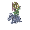
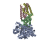
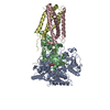
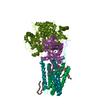
| Title | Plasticity of the quinone-binding site of the complex II homolog quinol:fumarate reductase | ||||||
 Components Components | (Fumarate reductase ... ) x 4 ) x 4 | ||||||
 Keywords Keywords | OXIDOREDUCTASE / Membrane Protein / Complex II homolog / FrdC-E29L | ||||||
| Function / homology |  Function and homology information Function and homology informationplasma membrane fumarate reductase complex / succinate dehydrogenase activity / fermentation / fumarate metabolic process / succinate dehydrogenase / anaerobic electron transport chain / succinate dehydrogenase (quinone) activity / anaerobic respiration / bacterial-type flagellum assembly / FAD binding ...plasma membrane fumarate reductase complex / succinate dehydrogenase activity / fermentation / fumarate metabolic process / succinate dehydrogenase / anaerobic electron transport chain / succinate dehydrogenase (quinone) activity / anaerobic respiration / bacterial-type flagellum assembly / FAD binding / flavin adenine dinucleotide binding / electron transfer activity / DNA damage response / membrane / plasma membrane / cytosolSimilarity search - Function | ||||||
| Biological species |  Escherichia coli (E. coli) Escherichia coli (E. coli) | ||||||
| Method | X-RAY DIFFRACTION / SYNCHROTRON / MOLECULAR REPLACEMENT / Resolution: 2.95 Å | ||||||
 Authors Authors | Singh, P.K. / Sarwar, M. / Maklashina, E. / Kotlyar, V. / Rajagukguk, S. / Tomasiak, T.M. / Cecchini, G. / Iverson, T.M. | ||||||
 Citation Citation | Journal: J.Biol.Chem. / Year: 2013 Title: Plasticity of the Quinone-binding Site of the Complex II Homolog Quinol:Fumarate Reductase. Authors: Singh, P.K. / Sarwar, M. / Maklashina, E. / Kotlyar, V. / Rajagukguk, S. / Tomasiak, T.M. / Cecchini, G. / Iverson, T.M. | ||||||
| History |
|
- Structure visualization
| Structure viewer | Molecule: MolmilJmol/JSmol |
|---|
- Downloads & links
-Download
| PDBx/mmCIF format | 4kx6.cif.gz | 831.8 KB | Display | PDBx/mmCIF format |
|---|---|---|---|---|
| PDB format | pdb4kx6.ent.gz | 692 KB | Display | PDB format |
| PDBx/mmJSON format | 4kx6.json.gz | Tree view | PDBx/mmJSON format | |
| Others |  Other downloads Other downloads |
-Validation report
| Arichive directory | https://data.pdbj.org/pub/pdb/validation_reports/kx/4kx6ftp://data.pdbj.org/pub/pdb/validation_reports/kx/4kx6 | HTTPS FTP |
|---|
-Related structure data
| Related structure data | |
|---|---|
| Similar structure data |


-Links
 PDBj
PDBj


- Assembly
| Deposited unit | 
| ||||||||
|---|---|---|---|---|---|---|---|---|---|
| 1 | 
| ||||||||
| 2 | 
| ||||||||
| Unit cell |
|
-Components
-Fumarate reductase ... , 4 types, 8 molecules AMBNCODP
| #1: Protein | Mass: 63477.707 Da / Num. of mol.: 2 Source method: isolated from a genetically manipulated source Source: (gene. exp.) Escherichia coli (E. coli) / Strain: K12 / Gene: frdA, b4154, JW4115 / Production host: Escherichia coli (E. coli) / References: UniProt: P00363, succinate dehydrogenase#2: Protein | Mass: 27021.885 Da / Num. of mol.: 2 Source method: isolated from a genetically manipulated source Source: (gene. exp.) Escherichia coli (E. coli) / Strain: K12 / MC4100 / BW2952 / Gene: frdB, BWG_3868 / Production host: Escherichia coli (E. coli) / References: UniProt: C5A1E7#3: Protein | / Fumarate reductase 15 kDa hydrophobic proteinMass: 14882.818 Da / Num. of mol.: 2 / Mutation: E29L Source method: isolated from a genetically manipulated source Source: (gene. exp.) Escherichia coli (E. coli) / Strain: ATCC 33849 / DSM 4235 / NCIB 12045 / K12 / DH1 / Gene: frdC, EcDH1_3838, ECDH1ME8569_4012 / Production host: Escherichia coli (E. coli) / References: UniProt: C9QU46#4: Protein | / Fumarate reductase 13 kDa hydrophobic proteinMass: 13118.870 Da / Num. of mol.: 2 Source method: isolated from a genetically manipulated source Source: (gene. exp.) Escherichia coli (E. coli) / Strain: K12 / Gene: frdD, b4151, JW4112 / Production host: Escherichia coli (E. coli) / References: UniProt: P0A8Q3 |
|---|
-Non-polymers , 5 types, 10 molecules 


| #5: Chemical | Flavin adenine dinucleotide Mass: 785.550 Da / Num. of mol.: 2 / Source method: obtained synthetically / Formula: C27H33N9O15P2 / Comment: FAD*YM Mass: 785.550 Da / Num. of mol.: 2 / Source method: obtained synthetically / Formula: C27H33N9O15P2 / Comment: FAD*YM#6: Chemical | Iron–sulfur cluster Mass: 175.820 Da / Num. of mol.: 2 / Source method: obtained synthetically / Formula: Fe2S2 Mass: 175.820 Da / Num. of mol.: 2 / Source method: obtained synthetically / Formula: Fe2S2#7: Chemical | Iron–sulfur cluster Mass: 295.795 Da / Num. of mol.: 2 / Source method: obtained synthetically / Formula: Fe3S4 Mass: 295.795 Da / Num. of mol.: 2 / Source method: obtained synthetically / Formula: Fe3S4#8: Chemical | Iron–sulfur cluster Mass: 351.640 Da / Num. of mol.: 2 / Source method: obtained synthetically / Formula: Fe4S4 Mass: 351.640 Da / Num. of mol.: 2 / Source method: obtained synthetically / Formula: Fe4S4#9: Chemical | Vitamin K2 Mass: 648.999 Da / Num. of mol.: 2 / Source method: obtained synthetically / Formula: C46H64O2 Mass: 648.999 Da / Num. of mol.: 2 / Source method: obtained synthetically / Formula: C46H64O2 |
|---|
-Experimental details
-Experiment
| Experiment | Method: X-RAY DIFFRACTION / Number of used crystals: 1 |
|---|
- Sample preparation
| Crystal | Density Matthews: 3.88 Å3/Da / Density % sol: 68.3 % |
|---|---|
| Crystal grow | Temperature: 295.15 K / Method: vapor diffusion, hanging drop / pH: 5.8 Details: 13%-15% PEG 5000 MME, 85mM-115mM Magnesium Acetate, 100mM Sodium Citrate, 0.01mM DTT, 0.1mM EDTA, pH 5.8, VAPOR DIFFUSION, HANGING DROP, temperature 295.15K |
-Data collection
| Diffraction | Mean temperature: 295.15 K | |||||||||||||||||||||||||||||||||
|---|---|---|---|---|---|---|---|---|---|---|---|---|---|---|---|---|---|---|---|---|---|---|---|---|---|---|---|---|---|---|---|---|---|---|
| Diffraction source | Source: SYNCHROTRON / Site: APS  / Beamline: 21-ID-G / Wavelength: 0.97856 Å / Beamline: 21-ID-G / Wavelength: 0.97856 Å | |||||||||||||||||||||||||||||||||
| Detector | Type: MAR scanner 300 mm plate / Detector: IMAGE PLATE / Date: Apr 7, 2012 | |||||||||||||||||||||||||||||||||
| Radiation | Monochromator: 3.0 cm undulator using two Laue diamond monochromators and one 2.5 m large offset monochromator Protocol: SINGLE WAVELENGTH / Monochromatic (M) / Laue (L): M / Scattering type: x-ray | |||||||||||||||||||||||||||||||||
| Radiation wavelength | Wavelength: 0.97856 Å / Relative weight: 1 | |||||||||||||||||||||||||||||||||
| Reflection | Resolution: 2.95→47.8 Å / Num. obs: 75158 / % possible obs: 95.7 % / Observed criterion σ(F): 33.2 / Observed criterion σ(I): 116.3 | |||||||||||||||||||||||||||||||||
| Reflection shell |
|
- Processing
| Software |
| ||||||||||||||||||||||||||||||||||||||||||||||||||||||||||||||||||||||||||||||||||||||||||||||||||||||||||||||||||||||||||||||||||||||||||||||||||||||||||||||||||||||||||||||||||||||
|---|---|---|---|---|---|---|---|---|---|---|---|---|---|---|---|---|---|---|---|---|---|---|---|---|---|---|---|---|---|---|---|---|---|---|---|---|---|---|---|---|---|---|---|---|---|---|---|---|---|---|---|---|---|---|---|---|---|---|---|---|---|---|---|---|---|---|---|---|---|---|---|---|---|---|---|---|---|---|---|---|---|---|---|---|---|---|---|---|---|---|---|---|---|---|---|---|---|---|---|---|---|---|---|---|---|---|---|---|---|---|---|---|---|---|---|---|---|---|---|---|---|---|---|---|---|---|---|---|---|---|---|---|---|---|---|---|---|---|---|---|---|---|---|---|---|---|---|---|---|---|---|---|---|---|---|---|---|---|---|---|---|---|---|---|---|---|---|---|---|---|---|---|---|---|---|---|---|---|---|---|---|---|---|
| Refinement | Method to determine structure: MOLECULAR REPLACEMENT / Resolution: 2.95→47.8 Å / Cor.coef. Fo:Fc: 0.905 / Cor.coef. Fo:Fc free: 0.88 / SU B: 37.421 / SU ML: 0.314 / Cross valid method: THROUGHOUT / ESU R: 1.164 / ESU R Free: 0.402 / Stereochemistry target values: MAXIMUM LIKELIHOOD / Details: HYDROGENS HAVE BEEN ADDED IN THE RIDING POSITIONS
| ||||||||||||||||||||||||||||||||||||||||||||||||||||||||||||||||||||||||||||||||||||||||||||||||||||||||||||||||||||||||||||||||||||||||||||||||||||||||||||||||||||||||||||||||||||||
| Solvent computation | Ion probe radii: 0.8 Å / Shrinkage radii: 0.8 Å / VDW probe radii: 1.2 Å / Solvent model: BABINET MODEL WITH MASK | ||||||||||||||||||||||||||||||||||||||||||||||||||||||||||||||||||||||||||||||||||||||||||||||||||||||||||||||||||||||||||||||||||||||||||||||||||||||||||||||||||||||||||||||||||||||
| Displacement parameters | Biso mean: 75.533 Å2
| ||||||||||||||||||||||||||||||||||||||||||||||||||||||||||||||||||||||||||||||||||||||||||||||||||||||||||||||||||||||||||||||||||||||||||||||||||||||||||||||||||||||||||||||||||||||
| Refinement step | Cycle: LAST / Resolution: 2.95→47.8 Å
| ||||||||||||||||||||||||||||||||||||||||||||||||||||||||||||||||||||||||||||||||||||||||||||||||||||||||||||||||||||||||||||||||||||||||||||||||||||||||||||||||||||||||||||||||||||||
| Refine LS restraints |
|