 Movie
Movie Controller


+ Open data

- Basic information
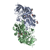
| Entry | Database: PDB / ID: 4kt1 | |||||||||
|---|---|---|---|---|---|---|---|---|---|---|
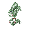
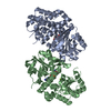
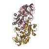
| Title | Complex of R-spondin 1 with LGR4 extracellular domain | |||||||||
 Components Components |
| |||||||||
 Keywords Keywords | HORMONE RECEPTOR/CELL ADHESION / R-spondin / LGR receptor / complex structure /  Wnt signaling / HORMONE RECEPTOR-CELL ADHESION complex Wnt signaling / HORMONE RECEPTOR-CELL ADHESION complex | |||||||||
| Function / homology |  Function and homology information Function and homology informationcell differentiation involved in metanephros development / metanephric glomerulus development / metanephric nephron tubule morphogenesis / : / epithelial cell proliferation involved in renal tubule morphogenesis / protein-hormone receptor activity / intestinal stem cell homeostasis / regulation of receptor internalization / negative regulation of toll-like receptor signaling pathway / positive regulation of branching involved in ureteric bud morphogenesis ...cell differentiation involved in metanephros development / metanephric glomerulus development / metanephric nephron tubule morphogenesis / : / epithelial cell proliferation involved in renal tubule morphogenesis / protein-hormone receptor activity / intestinal stem cell homeostasis / regulation of receptor internalization / negative regulation of toll-like receptor signaling pathway / positive regulation of branching involved in ureteric bud morphogenesis / male genitalia development / bone remodeling / G protein-coupled peptide receptor activity / digestive tract development / negative regulation of cold-induced thermogenesis / negative regulation of cytokine production / bone mineralization / positive regulation of Wnt signaling pathway / hair follicle development / Regulation of FZD by ubiquitination / G protein-coupled receptor binding / G protein-coupled receptor activity / circadian regulation of gene expression / adenylate cyclase-activating G protein-coupled receptor signaling pathway / Wnt signaling pathway / osteoblast differentiation / positive regulation of canonical Wnt signaling pathway / transmembrane signaling receptor activity / heparin binding / spermatogenesis / positive regulation of protein phosphorylation / signaling receptor binding / innate immune response / negative regulation of DNA-templated transcription / positive regulation of DNA-templated transcription / extracellular region / nucleus / plasma membraneSimilarity search - Function | |||||||||
| Biological species |  Homo sapiens (human) Homo sapiens (human) | |||||||||
| Method | X-RAY DIFFRACTION / SYNCHROTRON / MOLECULAR REPLACEMENT / Resolution: 2.497 Å | |||||||||
 Authors Authors | Wang, X.Q. / Wang, D.L. | |||||||||
 Citation Citation | Journal: Genes Dev. / Year: 2013 Title: Structural basis for R-spondin recognition by LGR4/5/6 receptors Authors: Wang, D.L. / Huang, B. / Zhang, S. / Yu, X. / Wu, W. / Wang, X.Q. | |||||||||
| History |
|
- Structure visualization
| Structure viewer | Molecule: MolmilJmol/JSmol |
|---|
- Downloads & links
-Download
| PDBx/mmCIF format | 4kt1.cif.gz | 231.8 KB | Display | PDBx/mmCIF format |
|---|---|---|---|---|
| PDB format | pdb4kt1.ent.gz | 184 KB | Display | PDB format |
| PDBx/mmJSON format | 4kt1.json.gz | Tree view | PDBx/mmJSON format | |
| Others |  Other downloads Other downloads |
-Validation report
| Arichive directory | https://data.pdbj.org/pub/pdb/validation_reports/kt/4kt1ftp://data.pdbj.org/pub/pdb/validation_reports/kt/4kt1 | HTTPS FTP |
|---|
-Related structure data
| Related structure data | 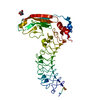 3zyoS S: Starting model for refinement |
|---|---|
| Similar structure data |


-Links
 PDBj
PDBj


- Assembly
| Deposited unit | 
| ||||||||
|---|---|---|---|---|---|---|---|---|---|
| 1 |
| ||||||||
| Unit cell |
|
-Components
| #1: Protein | Mass: 55438.344 Da / Num. of mol.: 1 / Fragment: UNP residues 26-502 Source method: isolated from a genetically manipulated source Source: (gene. exp.) Homo sapiens (human) / Gene: LGR4 / Plasmid: pFastBac / Production host:   Spodoptera frugiperda (fall armyworm) / References: UniProt: Q9BXB1 Spodoptera frugiperda (fall armyworm) / References: UniProt: Q9BXB1 |
|---|---|
| #2: Protein | Mass: 10080.942 Da / Num. of mol.: 1 / Fragment: UNP residues 39-128 Source method: isolated from a genetically manipulated source Source: (gene. exp.) Homo sapiens (human) / Gene: RSPO1 / Plasmid: pFastBac / Production host: Spodoptera frugiperda (fall armyworm) / References: UniProt: Q2MKA7 |
| #3: Polysaccharide | 2-acetamido-2-deoxy-beta-D-glucopyranose-(1-4)-2-acetamido-2-deoxy-beta-D-glucopyranose / Mass: 424.401 Da / Num. of mol.: 1 Source method: isolated from a genetically manipulated source |
| #4: Sugar | ChemComp-NAG / N-Acetylglucosamine  Type: D-saccharide, beta linking / Mass: 221.208 Da / Num. of mol.: 1 Type: D-saccharide, beta linking / Mass: 221.208 Da / Num. of mol.: 1Source method: isolated from a genetically manipulated source Formula: C8H15NO6 |
| #5: Water | ChemComp-HOH / Water Mass: 18.015 Da / Num. of mol.: 217 / Source method: isolated from a natural source / Formula: H2O Mass: 18.015 Da / Num. of mol.: 217 / Source method: isolated from a natural source / Formula: H2O |
-Experimental details
-Experiment
| Experiment | Method: X-RAY DIFFRACTION / Number of used crystals: 1 |
|---|
- Sample preparation
| Crystal | Density Matthews: 3.21 Å3/Da / Density % sol: 61.69 % |
|---|---|
| Crystal grow | Temperature: 291 K / Method: vapor diffusion, sitting drop / pH: 6 Details: PEG, pH 6.0, VAPOR DIFFUSION, SITTING DROP, temperature 291K |
-Data collection
| Diffraction | Mean temperature: 100 K |
|---|---|
| Diffraction source | Source: SYNCHROTRON / Site: SSRF  / Beamline: BL17U / Wavelength: 0.97 Å / Beamline: BL17U / Wavelength: 0.97 Å |
| Detector | Type: RIGAKU / Detector: IMAGE PLATE / Date: Jul 1, 2012 |
| Radiation | Monochromator: GRAPHITE / Protocol: SINGLE WAVELENGTH / Monochromatic (M) / Laue (L): M / Scattering type: x-ray |
| Radiation wavelength | Wavelength: 0.97 Å / Relative weight: 1 |
| Reflection | Resolution: 2.497→50 Å / Num. all: 28261 / Num. obs: 28261 / % possible obs: 99.9 % / Observed criterion σ(F): 1.96 / Observed criterion σ(I): 1.96 / Biso Wilson estimate: 35.06 Å2 |
| Reflection shell | Resolution: 2.5→2.56 Å / % possible all: 99.6 |
- Processing
| Software |
| ||||||||||||||||||||||||||||||||||||||||||||||||||||||||||||||||||||||||
|---|---|---|---|---|---|---|---|---|---|---|---|---|---|---|---|---|---|---|---|---|---|---|---|---|---|---|---|---|---|---|---|---|---|---|---|---|---|---|---|---|---|---|---|---|---|---|---|---|---|---|---|---|---|---|---|---|---|---|---|---|---|---|---|---|---|---|---|---|---|---|---|---|---|
| Refinement | Method to determine structure: MOLECULAR REPLACEMENT Starting model: 3ZYO Resolution: 2.497→24.671 Å / Occupancy max: 1 / Occupancy min: 1 / FOM work R set: 0.8596 / SU ML: 0.26 / σ(F): 1.96 / Phase error: 21.75 / Stereochemistry target values: ML
| ||||||||||||||||||||||||||||||||||||||||||||||||||||||||||||||||||||||||
| Solvent computation | Shrinkage radii: 0.9 Å / VDW probe radii: 1.11 Å / Solvent model: FLAT BULK SOLVENT MODEL | ||||||||||||||||||||||||||||||||||||||||||||||||||||||||||||||||||||||||
| Displacement parameters | Biso max: 118.64 Å2 / Biso mean: 41.7028 Å2 / Biso min: 17.17 Å2 | ||||||||||||||||||||||||||||||||||||||||||||||||||||||||||||||||||||||||
| Refinement step | Cycle: LAST / Resolution: 2.497→24.671 Å
| ||||||||||||||||||||||||||||||||||||||||||||||||||||||||||||||||||||||||
| Refine LS restraints |
| ||||||||||||||||||||||||||||||||||||||||||||||||||||||||||||||||||||||||
| LS refinement shell | Refine-ID: X-RAY DIFFRACTION / Total num. of bins used: 10 / % reflection obs: 100 %
| ||||||||||||||||||||||||||||||||||||||||||||||||||||||||||||||||||||||||
| Refinement TLS params. | S33: -0 Å ° / Method: refined / Refine-ID: X-RAY DIFFRACTION
| ||||||||||||||||||||||||||||||||||||||||||||||||||||||||||||||||||||||||
| Refinement TLS group |
|
