 Movie
Movie Controller

[English] 日本語
 Yorodumi
- PDB-3zyo: Crystal structure of the N-terminal leucine rich repeats and immu... -
+ Open data

- Basic information
| Entry | Database: PDB / ID: 3zyo | ||||||
|---|---|---|---|---|---|---|---|
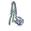
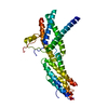
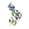
| Title | Crystal structure of the N-terminal leucine rich repeats and immunoglobulin domain of netrin-G ligand-3 | ||||||
 Components Components | LEUCINE-RICH REPEAT-CONTAINING PROTEIN 4B | ||||||
 Keywords Keywords |  CELL ADHESION / SYNAPSE CELL ADHESION / SYNAPSE | ||||||
| Function / homology |  Function and homology information Function and homology informationReceptor-type tyrosine-protein phosphatases / synaptic membrane adhesion / regulation of postsynaptic density assembly / cerebellar mossy fiber / positive regulation of synapse assembly / regulation of presynapse assembly / postsynaptic density membrane / presynaptic membrane / signaling receptor binding / glutamatergic synapse / plasma membraneSimilarity search - Function | ||||||
| Biological species |  MUS MUSCULUS (house mouse) MUS MUSCULUS (house mouse) | ||||||
| Method | X-RAY DIFFRACTION / SYNCHROTRON / MOLECULAR REPLACEMENT / Resolution: 3.1 Å | ||||||
 Authors Authors | Seiradake, E. / Coles, C.H. / Perestenko, P.V. / Harlos, K. / McIlhinney, R.A.J. / Aricescu, A.R. / Jones, E.Y. | ||||||
 Citation Citation | Journal: Embo J. / Year: 2011 Title: Structural Basis for Cell Surface Patterning Through Netring-Ngl Interactions. Authors: Seiradake, E. / Coles, C.H. / Perestenko, P.V. / Harlos, K. / Mcilhinney, R.A.J. / Aricescu, A.R. / Jones, E.Y. | ||||||
| History |
|
- Structure visualization
| Structure viewer | Molecule: MolmilJmol/JSmol |
|---|
- Downloads & links
-Download
| PDBx/mmCIF format | 3zyo.cif.gz | 172.1 KB | Display | PDBx/mmCIF format |
|---|---|---|---|---|
| PDB format | pdb3zyo.ent.gz | 138.3 KB | Display | PDB format |
| PDBx/mmJSON format | 3zyo.json.gz | Tree view | PDBx/mmJSON format | |
| Others |  Other downloads Other downloads |
-Validation report
| Arichive directory | https://data.pdbj.org/pub/pdb/validation_reports/zy/3zyoftp://data.pdbj.org/pub/pdb/validation_reports/zy/3zyo | HTTPS FTP |
|---|
-Related structure data
| Related structure data |  3zygC  3zyiC 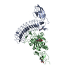 3zyjC 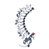 3zynSC S: Starting model for refinement C: citing same article ( |
|---|---|
| Similar structure data |


-Links
 PDBj
PDBj


- Assembly
| Deposited unit | 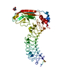
| ||||||||
|---|---|---|---|---|---|---|---|---|---|
| 1 |
| ||||||||
| Unit cell |
|
-Components
| #1: Protein | Mass: 46437.859 Da / Num. of mol.: 1 Fragment: N-TERMINAL LEUCINE RICH REPEATS AND IMMUNOGLOBULIN DOMAIN, RESIDUES 57-455 Source method: isolated from a genetically manipulated source Source: (gene. exp.) MUS MUSCULUS (house mouse) / Cell line (production host): HEK293S / Production host:  HOMO SAPIENS (human) / References: UniProt: P0C192 HOMO SAPIENS (human) / References: UniProt: P0C192 |
|---|---|
| #2: Chemical | ChemComp-ZN /   Mass: 65.409 Da / Num. of mol.: 1 / Source method: obtained synthetically / Formula: Zn Mass: 65.409 Da / Num. of mol.: 1 / Source method: obtained synthetically / Formula: Zn |
| #3: Sugar | ChemComp-NAG / N-Acetylglucosamine  Type: D-saccharide, beta linking / Mass: 221.208 Da / Num. of mol.: 1 Type: D-saccharide, beta linking / Mass: 221.208 Da / Num. of mol.: 1Source method: isolated from a genetically manipulated source Formula: C8H15NO6 |
-Experimental details
-Experiment
| Experiment | Method: X-RAY DIFFRACTION / Number of used crystals: 1 |
|---|
- Sample preparation
| Crystal | Density Matthews: 3.1 Å3/Da / Density % sol: 60 % / Description: NONE |
|---|---|
| Crystal grow | pH: 6.5 Details: 0.1 M NAH2PO4, 0.1 M KH2PO4, 2M NACL, 0.1 M MES, PH 6.5 |
-Data collection
| Diffraction | Mean temperature: 100 K |
|---|---|
| Diffraction source | Source: SYNCHROTRON / Site: Diamond  / Beamline: I03 / Wavelength: 1.0714 / Beamline: I03 / Wavelength: 1.0714 |
| Detector | Type: ADSC CCD / Detector: CCD / Date: Aug 5, 2009 |
| Radiation | Protocol: SINGLE WAVELENGTH / Monochromatic (M) / Laue (L): M / Scattering type: x-ray |
| Radiation wavelength | Wavelength: 1.0714 Å / Relative weight: 1 |
| Reflection | Resolution: 3.1→38.6 Å / Num. obs: 10743 / % possible obs: 99.9 % / Observed criterion σ(I): 2 / Redundancy: 7.3 % / Rmerge(I) obs: 0.13 / Net I/σ(I): 11.2 |
| Reflection shell | Resolution: 3.1→3.27 Å / Redundancy: 7.4 % / Rmerge(I) obs: 0.87 / Mean I/σ(I) obs: 2.6 / % possible all: 100 |
- Processing
| Software |
| ||||||||||||||||||||||||||||||||||||||||||||||||||||||||||||||||||||||||||||||||||||||||||||||||||||||||||||||||||||||||||||||||||||||||||||||||||||||||||||||||||||||||||||||||||||||
|---|---|---|---|---|---|---|---|---|---|---|---|---|---|---|---|---|---|---|---|---|---|---|---|---|---|---|---|---|---|---|---|---|---|---|---|---|---|---|---|---|---|---|---|---|---|---|---|---|---|---|---|---|---|---|---|---|---|---|---|---|---|---|---|---|---|---|---|---|---|---|---|---|---|---|---|---|---|---|---|---|---|---|---|---|---|---|---|---|---|---|---|---|---|---|---|---|---|---|---|---|---|---|---|---|---|---|---|---|---|---|---|---|---|---|---|---|---|---|---|---|---|---|---|---|---|---|---|---|---|---|---|---|---|---|---|---|---|---|---|---|---|---|---|---|---|---|---|---|---|---|---|---|---|---|---|---|---|---|---|---|---|---|---|---|---|---|---|---|---|---|---|---|---|---|---|---|---|---|---|---|---|---|---|
| Refinement | Method to determine structure: MOLECULAR REPLACEMENT Starting model: PDB ENTRY 3ZYN Resolution: 3.1→38.6 Å / Cor.coef. Fo:Fc: 0.931 / Cor.coef. Fo:Fc free: 0.889 / SU B: 49.006 / SU ML: 0.399 / Cross valid method: THROUGHOUT / ESU R Free: 0.469 / Stereochemistry target values: MAXIMUM LIKELIHOOD Details: HYDROGENS HAVE BEEN ADDED IN THE RIDING POSITIONS. CLEAR ELECTRON DENSITY FOR AMINO ACID RESIDUES Q68-A69 AND N335-C338 WAS NOT OBSERVED AND THESE ARE OMITTED FROM THE MODEL. DISORDERED ...Details: HYDROGENS HAVE BEEN ADDED IN THE RIDING POSITIONS. CLEAR ELECTRON DENSITY FOR AMINO ACID RESIDUES Q68-A69 AND N335-C338 WAS NOT OBSERVED AND THESE ARE OMITTED FROM THE MODEL. DISORDERED SIDECHAINS WERE MODELED STEREOCHEMICALLY
| ||||||||||||||||||||||||||||||||||||||||||||||||||||||||||||||||||||||||||||||||||||||||||||||||||||||||||||||||||||||||||||||||||||||||||||||||||||||||||||||||||||||||||||||||||||||
| Solvent computation | Ion probe radii: 0.8 Å / Shrinkage radii: 0.8 Å / VDW probe radii: 1.2 Å / Solvent model: MASK | ||||||||||||||||||||||||||||||||||||||||||||||||||||||||||||||||||||||||||||||||||||||||||||||||||||||||||||||||||||||||||||||||||||||||||||||||||||||||||||||||||||||||||||||||||||||
| Displacement parameters | Biso mean: 87.797 Å2
| ||||||||||||||||||||||||||||||||||||||||||||||||||||||||||||||||||||||||||||||||||||||||||||||||||||||||||||||||||||||||||||||||||||||||||||||||||||||||||||||||||||||||||||||||||||||
| Refinement step | Cycle: LAST / Resolution: 3.1→38.6 Å
| ||||||||||||||||||||||||||||||||||||||||||||||||||||||||||||||||||||||||||||||||||||||||||||||||||||||||||||||||||||||||||||||||||||||||||||||||||||||||||||||||||||||||||||||||||||||
| Refine LS restraints |
|