 Movie
Movie Controller


+ Open data

- Basic information
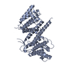
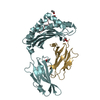
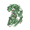
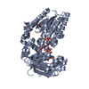
| Entry | Database: PDB / ID: 4krc | ||||||
|---|---|---|---|---|---|---|---|
| Title | Crystal Structure of Pho85-Pcl10-ATP-gamma-S Complex | ||||||
 Components Components |
| ||||||
 Keywords Keywords | Transferase/Signaling Protein /  Glycogen Synthesis / Glycogen Synthesis Regulation / Transferase-Signaling Protein complex Glycogen Synthesis / Glycogen Synthesis Regulation / Transferase-Signaling Protein complex | ||||||
| Function / homology |  Function and homology information Function and homology informationestablishment or maintenance of cytoskeleton polarity / Pho85-Pho80 CDK-cyclin complex / negative regulation of phosphate metabolic process / regulation of establishment or maintenance of cell polarity / negative regulation of calcium-mediated signaling / regulation of cell cycle phase transition / long-chain fatty acid metabolic process / positive regulation of phospholipid biosynthetic process / fungal-type cell wall organization / regulation of glycogen biosynthetic process ...establishment or maintenance of cytoskeleton polarity / Pho85-Pho80 CDK-cyclin complex / negative regulation of phosphate metabolic process / regulation of establishment or maintenance of cell polarity / negative regulation of calcium-mediated signaling / regulation of cell cycle phase transition / long-chain fatty acid metabolic process / positive regulation of phospholipid biosynthetic process / fungal-type cell wall organization / regulation of glycogen biosynthetic process / regulation of nucleocytoplasmic transport / negative regulation of glycogen biosynthetic process / cell cycle G1/S phase transition / cellular bud neck / cyclin-dependent protein serine/threonine kinase regulator activity / negative regulation of macroautophagy / glycogen metabolic process / regulation of cell division / lipid homeostasis / positive regulation of macroautophagy / regulation of lipid metabolic process / cyclin-dependent protein kinase holoenzyme complex / cyclin-dependent kinase / cyclin-dependent protein serine/threonine kinase activity / G1/S transition of mitotic cell cycle / regulation of protein stability / regulation of protein localization / regulation of cell cycle / protein kinase activity / phosphorylation / protein serine kinase activity / DNA damage response / regulation of DNA-templated transcription / regulation of transcription by RNA polymerase II / protein kinase binding / negative regulation of transcription by RNA polymerase II / ATP binding / nucleus / cytoplasmSimilarity search - Function | ||||||
| Biological species |  Saccharomyces cerevisiae (brewer's yeast) Saccharomyces cerevisiae (brewer's yeast) | ||||||
| Method | X-RAY DIFFRACTION / SYNCHROTRON / MOLECULAR REPLACEMENT / Resolution: 2.597 Å | ||||||
 Authors Authors | Quiocho, F.A. / Zheng, F. | ||||||
 Citation Citation | Journal: J.Biol.Chem. / Year: 2013 Title: New Structural Insights into Phosphorylation-free Mechanism for Full Cyclin-dependent Kinase (CDK)-Cyclin Activity and Substrate Recognition. Authors: Zheng, F. / Quiocho, F.A. | ||||||
| History |
|
- Structure visualization
| Structure viewer | Molecule: MolmilJmol/JSmol |
|---|
- Downloads & links
-Download
| PDBx/mmCIF format | 4krc.cif.gz | 195.5 KB | Display | PDBx/mmCIF format |
|---|---|---|---|---|
| PDB format | pdb4krc.ent.gz | 154.1 KB | Display | PDB format |
| PDBx/mmJSON format | 4krc.json.gz | Tree view | PDBx/mmJSON format | |
| Others |  Other downloads Other downloads |
-Validation report
| Arichive directory | https://data.pdbj.org/pub/pdb/validation_reports/kr/4krcftp://data.pdbj.org/pub/pdb/validation_reports/kr/4krc | HTTPS FTP |
|---|
-Related structure data


-Links
 PDBj
PDBj

- Assembly
| Deposited unit | 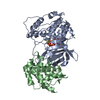
| ||||||||
|---|---|---|---|---|---|---|---|---|---|
| 1 |
| ||||||||
| Unit cell |
| ||||||||
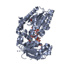
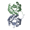
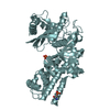
| Details | Heterodimer is composed of one molecular Pho85 and one molecular Pcl10 |
-Components
| #1: Protein | Mass: 36356.680 Da / Num. of mol.: 1 Source method: isolated from a genetically manipulated source Source: (gene. exp.) Saccharomyces cerevisiae (brewer's yeast)Strain: ATCC 204508 / S288c / Gene: P7102.18A, PHO85, SSG3, YPL031C / Plasmid: pQE60 / Production host:  Escherichia coli (E. coli) / Strain (production host): BL21 (DE3) / References: UniProt: P17157, cyclin-dependent kinase Escherichia coli (E. coli) / Strain (production host): BL21 (DE3) / References: UniProt: P17157, cyclin-dependent kinase |
|---|---|
| #2: Protein | Mass: 23752.602 Da / Num. of mol.: 1 / Fragment: UNP residues 227-433 Source method: isolated from a genetically manipulated source Source: (gene. exp.) Saccharomyces cerevisiae (brewer's yeast)Strain: ATCC 204508 / S288c / Gene: G2838, PCL10, YGL134W / Plasmid: pSBET / Production host: Escherichia coli (E. coli) / Strain (production host): BL21 (DE3) / References: UniProt: P53124 |
| #3: Chemical | ChemComp-MG /   Mass: 24.305 Da / Num. of mol.: 1 / Source method: obtained synthetically / Formula: Mg Mass: 24.305 Da / Num. of mol.: 1 / Source method: obtained synthetically / Formula: Mg |
| #4: Chemical | ChemComp-AGS /   Mass: 523.247 Da / Num. of mol.: 1 / Source method: obtained synthetically / Formula: C10H16N5O12P3S / Comment: ATP-gamma-S, energy-carrying molecule analogue*YM Mass: 523.247 Da / Num. of mol.: 1 / Source method: obtained synthetically / Formula: C10H16N5O12P3S / Comment: ATP-gamma-S, energy-carrying molecule analogue*YM |
| #5: Water | ChemComp-HOH / Water Mass: 18.015 Da / Num. of mol.: 23 / Source method: isolated from a natural source / Formula: H2O Mass: 18.015 Da / Num. of mol.: 23 / Source method: isolated from a natural source / Formula: H2O |
-Experimental details
-Experiment
| Experiment | Method: X-RAY DIFFRACTION / Number of used crystals: 1 |
|---|
- Sample preparation
| Crystal | Density Matthews: 2.15 Å3/Da / Density % sol: 42.82 % |
|---|---|
| Crystal grow | Temperature: 298 K / Method: vapor diffusion, hanging drop / pH: 6.4 Details: 22-25% Polyethylene glycol 5000 monomethyl ether (PEG 5K MME), 0.1 M 2-morpholinoethanesulfonic acid (MES), pH 6.4, VAPOR DIFFUSION, HANGING DROP, temperature 298K |
-Data collection
| Diffraction | Mean temperature: 100 K |
|---|---|
| Diffraction source | Source: SYNCHROTRON / Site: APS  / Beamline: 19-ID / Wavelength: 0.97925 Å / Beamline: 19-ID / Wavelength: 0.97925 Å |
| Detector | Type: ADSC QUANTUM 315 / Detector: CCD / Date: Apr 5, 2007 |
| Radiation | Monochromator: SAGITALLY FOCUSED SI(111) / Protocol: SINGLE WAVELENGTH / Monochromatic (M) / Laue (L): M / Scattering type: x-ray |
| Radiation wavelength | Wavelength: 0.97925 Å / Relative weight: 1 |
| Reflection | Resolution: 2.597→39.441 Å / Num. all: 15921 / Num. obs: 15166 / % possible obs: 95.26 % / Observed criterion σ(F): 1 / Observed criterion σ(I): 1 |
| Reflection shell | Resolution: 2.6→2.69 Å / Redundancy: 2.9 % / Rmerge(I) obs: 0.41 / % possible all: 73.4 |
- Processing
| Software |
| ||||||||||||||||||||||||||||||||||||||||||||||||||||||||||||||||||||||||
|---|---|---|---|---|---|---|---|---|---|---|---|---|---|---|---|---|---|---|---|---|---|---|---|---|---|---|---|---|---|---|---|---|---|---|---|---|---|---|---|---|---|---|---|---|---|---|---|---|---|---|---|---|---|---|---|---|---|---|---|---|---|---|---|---|---|---|---|---|---|---|---|---|---|
| Refinement | Method to determine structure: MOLECULAR REPLACEMENT / Resolution: 2.597→39.441 Å / SU ML: 0.4 / Cross valid method: THROUGHOUT / σ(F): 1.36 / Phase error: 30.77 / Stereochemistry target values: ML
| ||||||||||||||||||||||||||||||||||||||||||||||||||||||||||||||||||||||||
| Solvent computation | Shrinkage radii: 0.9 Å / VDW probe radii: 1.11 Å / Solvent model: FLAT BULK SOLVENT MODEL / Bsol: 81.393 Å2 / ksol: 0.345 e/Å3 | ||||||||||||||||||||||||||||||||||||||||||||||||||||||||||||||||||||||||
| Displacement parameters |
| ||||||||||||||||||||||||||||||||||||||||||||||||||||||||||||||||||||||||
| Refinement step | Cycle: LAST / Resolution: 2.597→39.441 Å
| ||||||||||||||||||||||||||||||||||||||||||||||||||||||||||||||||||||||||
| Refine LS restraints |
| ||||||||||||||||||||||||||||||||||||||||||||||||||||||||||||||||||||||||
| LS refinement shell |
| ||||||||||||||||||||||||||||||||||||||||||||||||||||||||||||||||||||||||
| Refinement TLS params. | S33: -0 Å ° / Method: refined / Refine-ID: X-RAY DIFFRACTION
| ||||||||||||||||||||||||||||||||||||||||||||||||||||||||||||||||||||||||
| Refinement TLS group |
|
