 Movie
Movie Controller

[English] 日本語
 Yorodumi
- PDB-1wv2: Crystal structure of thiamine biosynthesis protein from Pseudomon... -
+ Open data

- Basic information
| Entry | Database: PDB / ID: 1wv2 | ||||||
|---|---|---|---|---|---|---|---|
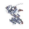
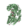
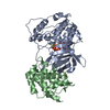
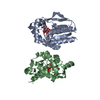
| Title | Crystal structure of thiamine biosynthesis protein from Pseudomonas Aeruginosa | ||||||
 Components Components | Thiazole biosynthesis protein thiG | ||||||
 Keywords Keywords |  BIOSYNTHETIC PROTEIN / Structural genomics / PROTEIN STRUCTURE INITIATIVE / PSI / New York SGX Research Center for Structural Genomics / NYSGXRC target T1779 / thiamine biosynthesis / TIM barrel BIOSYNTHETIC PROTEIN / Structural genomics / PROTEIN STRUCTURE INITIATIVE / PSI / New York SGX Research Center for Structural Genomics / NYSGXRC target T1779 / thiamine biosynthesis / TIM barrel | ||||||
| Function / homology |  Function and homology informationthiazole synthase / sulfurtransferase activity / thiamine diphosphate biosynthetic process / thiamine biosynthetic process / cytoplasm Function and homology informationthiazole synthase / sulfurtransferase activity / thiamine diphosphate biosynthetic process / thiamine biosynthetic process / cytoplasmSimilarity search - Function | ||||||
| Biological species |   Pseudomonas aeruginosa (bacteria) Pseudomonas aeruginosa (bacteria) | ||||||
| Method | X-RAY DIFFRACTION / SYNCHROTRON / MAD / Resolution: 2.9 Å | ||||||
 Authors Authors | Fedorov, A.A. / Fedorov, E.V. / Almo, S.C. / Burley, S.K. / New York SGX Research Center for Structural Genomics (NYSGXRC) | ||||||
 Citation Citation | Journal: To be Published Title: Crystal structure of thiamine biosynthesis protein from Pseudomonas Aeruginosa Authors: Fedorov, A.A. / Fedorov, E.V. / Almo, S.C. | ||||||
| History |
|
- Structure visualization
| Structure viewer | Molecule: MolmilJmol/JSmol |
|---|
- Downloads & links
-Download
| PDBx/mmCIF format | 1wv2.cif.gz | 94.6 KB | Display | PDBx/mmCIF format |
|---|---|---|---|---|
| PDB format | pdb1wv2.ent.gz | 72.8 KB | Display | PDB format |
| PDBx/mmJSON format | 1wv2.json.gz | Tree view | PDBx/mmJSON format | |
| Others |  Other downloads Other downloads |
-Validation report
| Arichive directory | https://data.pdbj.org/pub/pdb/validation_reports/wv/1wv2ftp://data.pdbj.org/pub/pdb/validation_reports/wv/1wv2 | HTTPS FTP |
|---|
-Related structure data
| Similar structure data | |
|---|---|
| Other databases |


-Links
 PDBj
PDBj- Assembly
| Deposited unit | 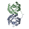
| ||||||||
|---|---|---|---|---|---|---|---|---|---|
| 1 |
| ||||||||
| 2 | 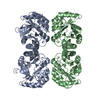
| ||||||||
| Unit cell |
|
-Components
| #1: Protein | Mass: 28262.658 Da / Num. of mol.: 2 Source method: isolated from a genetically manipulated source Source: (gene. exp.) Pseudomonas aeruginosa (bacteria) / Production host: Escherichia coli (E. coli) / References: UniProt: Q9I6B4 |
|---|
-Experimental details
-Experiment
| Experiment | Method: X-RAY DIFFRACTION / Number of used crystals: 1 |
|---|
- Sample preparation
| Crystal | Density Matthews: 2.21 Å3/Da / Density % sol: 42.2 % |
|---|---|
| Crystal grow | Temperature: 293 K / Method: vapor diffusion / pH: 4.5 Details: Sodium Chloride, Sodium acetate, pH 4.5, VAPOR DIFFUSION, temperature 293.0K |
-Data collection
| Diffraction | Mean temperature: 100 K | |||||||||||||||
|---|---|---|---|---|---|---|---|---|---|---|---|---|---|---|---|---|
| Diffraction source | Source: SYNCHROTRON / Site: NSLS  / Beamline: X9A / Wavelength: 0.979, 0.97934, 0.97911, 0.97166 / Beamline: X9A / Wavelength: 0.979, 0.97934, 0.97911, 0.97166 | |||||||||||||||
| Detector | Type: MARRESEARCH / Detector: CCD / Date: Sep 26, 2004 | |||||||||||||||
| Radiation | Protocol: MAD / Monochromatic (M) / Laue (L): M / Scattering type: x-ray | |||||||||||||||
| Radiation wavelength |
| |||||||||||||||
| Reflection | Resolution: 2.9→25 Å / Num. all: 11974 / Num. obs: 11974 / % possible obs: 98.3 % / Observed criterion σ(F): 0 / Observed criterion σ(I): 0 | |||||||||||||||
| Reflection shell | Resolution: 2.9→3 Å / % possible all: 97.1 |
- Processing
| Software |
| ||||||||||||||||||||
|---|---|---|---|---|---|---|---|---|---|---|---|---|---|---|---|---|---|---|---|---|---|
| Refinement | Method to determine structure: MAD / Resolution: 2.9→25 Å / σ(F): 0 / Stereochemistry target values: Engh & Huber
| ||||||||||||||||||||
| Refinement step | Cycle: LAST / Resolution: 2.9→25 Å
| ||||||||||||||||||||
| Refine LS restraints |
|
