 Movie
Movie Controller

[English] 日本語
 Yorodumi
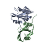
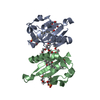
- PDB-4kr1: Crystal structure of the kinetechore protein Iml3 from budding yeast -
+ Open data

- Basic information
| Entry | Database: PDB / ID: 4kr1 | ||||||
|---|---|---|---|---|---|---|---|
| Title | Crystal structure of the kinetechore protein Iml3 from budding yeast | ||||||
 Components Components | Central kinetochore subunit IML3 | ||||||
 Keywords Keywords |  CELL CYCLE / chromosome segregation / kinetochore protein CELL CYCLE / chromosome segregation / kinetochore protein | ||||||
| Function / homology |  Function and homology information Function and homology informationmaintenance of meiotic sister chromatid cohesion / meiotic sister chromatid segregation / establishment of meiotic sister chromatid cohesion / ascospore formation / attachment of spindle microtubules to kinetochore / outer kinetochore / protein localization to chromosome, centromeric region / establishment of mitotic sister chromatid cohesion / kinetochore / cell division / nucleusSimilarity search - Function | ||||||
| Biological species |  Saccharomyces cerevisiae (brewer's yeast) Saccharomyces cerevisiae (brewer's yeast) | ||||||
| Method | X-RAY DIFFRACTION / SYNCHROTRON / SAD / Resolution: 2.5 Å | ||||||
 Authors Authors | Tao, Y. / Guo, Q. / Teng, M. | ||||||
 Citation Citation | Journal: Acta Crystallogr.,Sect.D / Year: 2013 Title: Structural insights into the role of the Chl4-Iml3 complex in kinetochore assembly Authors: Guo, Q. / Tao, Y. / Liu, H. / Teng, M. / Li, X. | ||||||
| History |
|
- Structure visualization
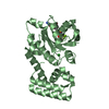
| Structure viewer | Molecule: MolmilJmol/JSmol |
|---|
- Downloads & links
-Download
| PDBx/mmCIF format | 4kr1.cif.gz | 94.1 KB | Display | PDBx/mmCIF format |
|---|---|---|---|---|
| PDB format | pdb4kr1.ent.gz | 76.7 KB | Display | PDB format |
| PDBx/mmJSON format | 4kr1.json.gz | Tree view | PDBx/mmJSON format | |
| Others |  Other downloads Other downloads |
-Validation report
| Arichive directory | https://data.pdbj.org/pub/pdb/validation_reports/kr/4kr1ftp://data.pdbj.org/pub/pdb/validation_reports/kr/4kr1 | HTTPS FTP |
|---|
-Related structure data
| Similar structure data |
|---|


-Links
 PDBj
PDBj- Assembly
| Deposited unit | 
| ||||||||
|---|---|---|---|---|---|---|---|---|---|
| 1 |
| ||||||||
| Unit cell |
|
-Components
| #1: Protein | Mass: 28659.406 Da / Num. of mol.: 1 Source method: isolated from a genetically manipulated source Source: (gene. exp.) Saccharomyces cerevisiae (brewer's yeast)Strain: S288c / Gene: IML3, MCM19, YBR107C, YBR0836 / Production host:  Escherichia coli (E. coli) / References: UniProt: P38265 Escherichia coli (E. coli) / References: UniProt: P38265 |
|---|---|
| #2: Water | ChemComp-HOH / Water Mass: 18.015 Da / Num. of mol.: 22 / Source method: isolated from a natural source / Formula: H2O Mass: 18.015 Da / Num. of mol.: 22 / Source method: isolated from a natural source / Formula: H2O |
-Experimental details
-Experiment
| Experiment | Method: X-RAY DIFFRACTION / Number of used crystals: 1 |
|---|
- Sample preparation
| Crystal | Density Matthews: 2.54 Å3/Da / Density % sol: 51.49 % |
|---|---|
| Crystal grow | Temperature: 285 K / Method: vapor diffusion, hanging drop / pH: 7 Details: 3M Sodium formate, 100mM Cesium chloride, 3% PEG 4000, pH 7.0, VAPOR DIFFUSION, HANGING DROP, temperature 285K |
-Data collection
| Diffraction | Mean temperature: 100 K |
|---|---|
| Diffraction source | Source: SYNCHROTRON / Site: SSRF  / Beamline: BL17U / Wavelength: 0.9794 Å / Beamline: BL17U / Wavelength: 0.9794 Å |
| Detector | Type: MARMOSAIC 225 mm CCD / Detector: CCD / Date: Jan 20, 2010 |
| Radiation | Monochromator: SAGITALLY FOCUSED Si(111) / Protocol: SINGLE WAVELENGTH / Monochromatic (M) / Laue (L): M / Scattering type: x-ray |
| Radiation wavelength | Wavelength: 0.9794 Å / Relative weight: 1 |
| Reflection | Resolution: 2.5→50 Å / Num. obs: 11039 / % possible obs: 99.9 % / Redundancy: 20.2 % / Biso Wilson estimate: 50.66 Å2 / Rmerge(I) obs: 0.101 / Net I/σ(I): 35.9 |
| Reflection shell | Resolution: 2.5→2.54 Å / Redundancy: 20.6 % / Rmerge(I) obs: 0.686 / Mean I/σ(I) obs: 5.5 / % possible all: 100 |
- Processing
| Software |
| |||||||||||||||||||||||||||||||||||||||||||||||||||||||||||||||||||||||||||||||||||||
|---|---|---|---|---|---|---|---|---|---|---|---|---|---|---|---|---|---|---|---|---|---|---|---|---|---|---|---|---|---|---|---|---|---|---|---|---|---|---|---|---|---|---|---|---|---|---|---|---|---|---|---|---|---|---|---|---|---|---|---|---|---|---|---|---|---|---|---|---|---|---|---|---|---|---|---|---|---|---|---|---|---|---|---|---|---|---|
| Refinement | Method to determine structure: SAD / Resolution: 2.5→47.21 Å / Cor.coef. Fo:Fc: 0.93 / Cor.coef. Fo:Fc free: 0.935 / Occupancy max: 1 / Occupancy min: 1 / SU B: 20.975 / SU ML: 0.205 / Cross valid method: THROUGHOUT / σ(F): 0 / ESU R: 0.414 / ESU R Free: 0.27 / Stereochemistry target values: MAXIMUM LIKELIHOOD Details: HYDROGENS HAVE BEEN ADDED IN THE RIDING POSITIONS U VALUES: WITH TLS ADDED
| |||||||||||||||||||||||||||||||||||||||||||||||||||||||||||||||||||||||||||||||||||||
| Solvent computation | Ion probe radii: 0.8 Å / Shrinkage radii: 0.8 Å / VDW probe radii: 1.4 Å / Solvent model: MASK | |||||||||||||||||||||||||||||||||||||||||||||||||||||||||||||||||||||||||||||||||||||
| Displacement parameters | Biso max: 145.54 Å2 / Biso mean: 49.0334 Å2 / Biso min: 25.72 Å2
| |||||||||||||||||||||||||||||||||||||||||||||||||||||||||||||||||||||||||||||||||||||
| Refinement step | Cycle: LAST / Resolution: 2.5→47.21 Å
| |||||||||||||||||||||||||||||||||||||||||||||||||||||||||||||||||||||||||||||||||||||
| Refine LS restraints |
| |||||||||||||||||||||||||||||||||||||||||||||||||||||||||||||||||||||||||||||||||||||
| LS refinement shell | Resolution: 2.501→2.566 Å / Total num. of bins used: 20
| |||||||||||||||||||||||||||||||||||||||||||||||||||||||||||||||||||||||||||||||||||||
| Refinement TLS params. | Method: refined / Refine-ID: X-RAY DIFFRACTION
| |||||||||||||||||||||||||||||||||||||||||||||||||||||||||||||||||||||||||||||||||||||
| Refinement TLS group |
|
