 Movie
Movie Controller


+ Open data

- Basic information
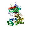
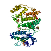
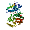
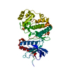
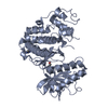
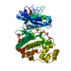
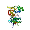
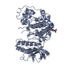
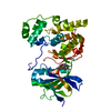
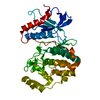
| Entry | Database: PDB / ID: 4kkh | ||||||
|---|---|---|---|---|---|---|---|
| Title | The crystal structure of inhibitor-bound JNK3 | ||||||
 Components Components | Mitogen-activated protein kinase 10 | ||||||
 Keywords Keywords | TRANSFERASE/TRANSFERASE INHIBITOR /  kinase domain / kinase / c-jun / TRANSFERASE-TRANSFERASE INHIBITOR complex kinase domain / kinase / c-jun / TRANSFERASE-TRANSFERASE INHIBITOR complex | ||||||
| Function / homology |  Function and homology information Function and homology informationJUN kinase activity / Activation of the AP-1 family of transcription factors / Fc-epsilon receptor signaling pathway / MAP kinase kinase activity / response to light stimulus / mitogen-activated protein kinase / JNK cascade / JNK (c-Jun kinases) phosphorylation and activation mediated by activated human TAK1 / FCERI mediated MAPK activation / regulation of circadian rhythm ...JUN kinase activity / Activation of the AP-1 family of transcription factors / Fc-epsilon receptor signaling pathway / MAP kinase kinase activity / response to light stimulus / mitogen-activated protein kinase / JNK cascade / JNK (c-Jun kinases) phosphorylation and activation mediated by activated human TAK1 / FCERI mediated MAPK activation / regulation of circadian rhythm / cellular senescence / rhythmic process / Oxidative Stress Induced Senescence / protein phosphorylation / protein serine kinase activity / signal transduction / mitochondrion / nucleoplasm / ATP binding / nucleus / plasma membrane / cytosol / cytoplasmSimilarity search - Function | ||||||
| Biological species |  Homo sapiens (human) Homo sapiens (human) | ||||||
| Method | X-RAY DIFFRACTION / SYNCHROTRON / MOLECULAR REPLACEMENT / Resolution: 2 Å | ||||||
 Authors Authors | Han, B.G. / Shim, M.B. / Ahn, H.C. | ||||||
 Citation Citation | Journal: To be Published Title: The crystal structure of AMP, apo, and inhibitor-bound JNK3 Authors: Han, B.G. / Shim, M.B. / Ahn, H.C. | ||||||
| History |
|
- Structure visualization
| Structure viewer | Molecule: MolmilJmol/JSmol |
|---|
- Downloads & links
-Download
| PDBx/mmCIF format | 4kkh.cif.gz | 88.2 KB | Display | PDBx/mmCIF format |
|---|---|---|---|---|
| PDB format | pdb4kkh.ent.gz | 65.6 KB | Display | PDB format |
| PDBx/mmJSON format | 4kkh.json.gz | Tree view | PDBx/mmJSON format | |
| Others |  Other downloads Other downloads |
-Validation report
| Arichive directory | https://data.pdbj.org/pub/pdb/validation_reports/kk/4kkhftp://data.pdbj.org/pub/pdb/validation_reports/kk/4kkh | HTTPS FTP |
|---|
-Related structure data
| Related structure data | |
|---|---|
| Similar structure data |


-Links
 PDBj
PDBj


- Assembly
| Deposited unit | 
| ||||||||
|---|---|---|---|---|---|---|---|---|---|
| 1 |
| ||||||||
| Unit cell |
|
-Components
| #1: Protein | Mass: 42116.727 Da / Num. of mol.: 1 Source method: isolated from a genetically manipulated source Source: (gene. exp.) Homo sapiens (human) / Gene: MAPK10, JNK3, JNK3A, PRKM10, SAPK1B / Production host:  Escherichia coli (E. coli) Escherichia coli (E. coli)References: UniProt: P53779, mitogen-activated protein kinase |
|---|---|
| #2: Chemical | ChemComp-1RQ /   Mass: 504.582 Da / Num. of mol.: 1 / Source method: obtained synthetically / Formula: C30H28N6O2 Mass: 504.582 Da / Num. of mol.: 1 / Source method: obtained synthetically / Formula: C30H28N6O2 |
| #3: Water | ChemComp-HOH / Water Mass: 18.015 Da / Num. of mol.: 155 / Source method: isolated from a natural source / Formula: H2O Mass: 18.015 Da / Num. of mol.: 155 / Source method: isolated from a natural source / Formula: H2O |
-Experimental details
-Experiment
| Experiment | Method: X-RAY DIFFRACTION / Number of used crystals: 1 |
|---|
- Sample preparation
| Crystal | Density Matthews: 2.38 Å3/Da / Density % sol: 48.27 % |
|---|---|
| Crystal grow | Temperature: 277 K / Method: vapor diffusion, hanging drop / pH: 7.25 Details: 16% PEG MME 550, 10% Ethylene glycol, 0.1 M Hepes, 10 mM TCEP, pH 7.25, VAPOR DIFFUSION, HANGING DROP, temperature 277K |
-Data collection
| Diffraction | Mean temperature: 100 K |
|---|---|
| Diffraction source | Source: SYNCHROTRON / Site: PAL/PLS  / Beamline: 5C (4A) / Beamline: 5C (4A) |
| Detector | Type: ADSC QUANTUM 315 / Detector: CCD / Date: Nov 23, 2012 |
| Radiation | Protocol: SINGLE WAVELENGTH / Monochromatic (M) / Laue (L): M / Scattering type: x-ray |
| Radiation wavelength | Relative weight: 1 |
| Reflection | Resolution: 2→37.45 Å / Num. obs: 26471 |
- Processing
| Software |
| |||||||||||||||||||||||||||||||||||||||||||||
|---|---|---|---|---|---|---|---|---|---|---|---|---|---|---|---|---|---|---|---|---|---|---|---|---|---|---|---|---|---|---|---|---|---|---|---|---|---|---|---|---|---|---|---|---|---|---|
| Refinement | Method to determine structure: MOLECULAR REPLACEMENT / Resolution: 2→37.45 Å / Cor.coef. Fo:Fc: 0.947 / Cor.coef. Fo:Fc free: 0.917 / SU B: 5.212 / SU ML: 0.144 / Cross valid method: THROUGHOUT / ESU R: 0.215 / ESU R Free: 0.191 / Stereochemistry target values: MAXIMUM LIKELIHOOD / Details: HYDROGENS HAVE BEEN USED IF PRESENT IN THE INPUT
| |||||||||||||||||||||||||||||||||||||||||||||
| Solvent computation | Ion probe radii: 0.8 Å / Shrinkage radii: 0.8 Å / VDW probe radii: 1.2 Å / Solvent model: MASK | |||||||||||||||||||||||||||||||||||||||||||||
| Displacement parameters | Biso mean: 43.869 Å2
| |||||||||||||||||||||||||||||||||||||||||||||
| Refinement step | Cycle: LAST / Resolution: 2→37.45 Å
| |||||||||||||||||||||||||||||||||||||||||||||
| Refine LS restraints |
| |||||||||||||||||||||||||||||||||||||||||||||
| LS refinement shell | Resolution: 2→2.051 Å / Total num. of bins used: 20
|
