 Movie
Movie Controller


+ Open data

- Basic information
| Entry | Database: PDB / ID: 4kh3 | ||||||
|---|---|---|---|---|---|---|---|
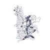
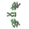
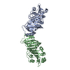
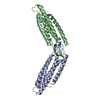
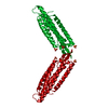
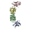
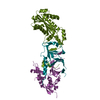
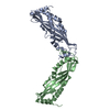
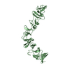
| Title | Structure of a bacterial self-associating protein | ||||||
 Components Components | Antigen 43 | ||||||
 Keywords Keywords | IMMUNE SYSTEM / Self-associating protein / Uropathogenic Escherichia coli / AIDA-I type autotransporter / Ag43 / aggregation / biofilm | ||||||
| Function / homology |  Function and homology information Function and homology information | ||||||
| Biological species |  Escherichia coli (E. coli) Escherichia coli (E. coli) | ||||||
| Method | X-RAY DIFFRACTION / SYNCHROTRON / SAD / Resolution: 2.5 Å | ||||||
 Authors Authors | Heras, B. / Gee, C.L. / Schembri, M.A. / Totsika, M. | ||||||
 Citation Citation | Journal: Proc.Natl.Acad.Sci.USA / Year: 2014 Title: The antigen 43 structure reveals a molecular Velcro-like mechanism of autotransporter-mediated bacterial clumping. Authors: Heras, B. / Totsika, M. / Peters, K.M. / Paxman, J.J. / Gee, C.L. / Jarrott, R.J. / Perugini, M.A. / Whitten, A.E. / Schembri, M.A. | ||||||
| History |
|
- Structure visualization
| Structure viewer | Molecule: MolmilJmol/JSmol |
|---|
- Downloads & links
-Download
| PDBx/mmCIF format | 4kh3.cif.gz | 107 KB | Display | PDBx/mmCIF format |
|---|---|---|---|---|
| PDB format | pdb4kh3.ent.gz | 81.8 KB | Display | PDB format |
| PDBx/mmJSON format | 4kh3.json.gz | Tree view | PDBx/mmJSON format | |
| Others |  Other downloads Other downloads |
-Validation report
| Arichive directory | https://data.pdbj.org/pub/pdb/validation_reports/kh/4kh3ftp://data.pdbj.org/pub/pdb/validation_reports/kh/4kh3 | HTTPS FTP |
|---|
-Related structure data
| Related structure data | |
|---|---|
| Similar structure data |


-Links
 PDBj
PDBj- Assembly
| Deposited unit | 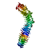
| |||||||||
|---|---|---|---|---|---|---|---|---|---|---|
| 1 |
| |||||||||
| Unit cell |
| |||||||||
| Components on special symmetry positions |
|
-Components
| #1: Protein | Mass: 50169.238 Da / Num. of mol.: 1 / Fragment: alpha domain, UNP RESIDUES 55-554 Source method: isolated from a genetically manipulated source Source: (gene. exp.) Escherichia coli (E. coli) / Strain: CFT073 / Gene: agn43, c3655 / Plasmid: pETLIC / Production host: Escherichia coli (E. coli) / Strain (production host): BL21(DE3) pLysS / References: UniProt: Q8CVR0, UniProt: A0A0H2VAW5*PLUS | ||
|---|---|---|---|
| #2: Chemical | ChemComp-MLI / Malonic acid  Mass: 102.046 Da / Num. of mol.: 7 / Source method: obtained synthetically / Formula: C3H2O4 Mass: 102.046 Da / Num. of mol.: 7 / Source method: obtained synthetically / Formula: C3H2O4#3: Water | ChemComp-HOH / | Water Mass: 18.015 Da / Num. of mol.: 306 / Source method: isolated from a natural source / Formula: H2O Mass: 18.015 Da / Num. of mol.: 306 / Source method: isolated from a natural source / Formula: H2O |
-Experimental details
-Experiment
| Experiment | Method: X-RAY DIFFRACTION / Number of used crystals: 2 |
|---|
- Sample preparation
| Crystal | Density Matthews: 4.13 Å3/Da / Density % sol: 70.25 % |
|---|---|
| Crystal grow | Temperature: 291 K / Method: vapor diffusion / pH: 4 Details: 2.8M sodium malonate pH 4, 10mM ATP, VAPOR DIFFUSION, temperature 291K |
-Data collection
| Diffraction |
| ||||||||||||||||||
|---|---|---|---|---|---|---|---|---|---|---|---|---|---|---|---|---|---|---|---|
| Diffraction source |
| ||||||||||||||||||
| Detector |
| ||||||||||||||||||
| Radiation |
| ||||||||||||||||||
| Radiation wavelength |
| ||||||||||||||||||
| Reflection | Resolution: 2.5→19.7 Å / Num. all: 29768 / Num. obs: 29768 / % possible obs: 99.8 % / Redundancy: 14.2 % / Rmerge(I) obs: 0.124 | ||||||||||||||||||
| Reflection shell | Resolution: 2.5→2.64 Å / Redundancy: 13.9 % / Rmerge(I) obs: 0.575 / Mean I/σ(I) obs: 4.6 / Num. unique all: 4278 / % possible all: 99.9 |

- Processing
| Software |
| ||||||||||||||||||||||||||||||||||||||||||||||||||||||||||||||||||||||||||||||||||||||||||||||||||||||||||||||||||||||||||||||
|---|---|---|---|---|---|---|---|---|---|---|---|---|---|---|---|---|---|---|---|---|---|---|---|---|---|---|---|---|---|---|---|---|---|---|---|---|---|---|---|---|---|---|---|---|---|---|---|---|---|---|---|---|---|---|---|---|---|---|---|---|---|---|---|---|---|---|---|---|---|---|---|---|---|---|---|---|---|---|---|---|---|---|---|---|---|---|---|---|---|---|---|---|---|---|---|---|---|---|---|---|---|---|---|---|---|---|---|---|---|---|---|---|---|---|---|---|---|---|---|---|---|---|---|---|---|---|---|
| Refinement | Method to determine structure: SAD / Resolution: 2.5→19.692 Å / SU ML: 0.32 / σ(F): 1.34 / Phase error: 19.59 / Stereochemistry target values: ML
| ||||||||||||||||||||||||||||||||||||||||||||||||||||||||||||||||||||||||||||||||||||||||||||||||||||||||||||||||||||||||||||||
| Solvent computation | Shrinkage radii: 1.11 Å / VDW probe radii: 1.3 Å / Solvent model: FLAT BULK SOLVENT MODEL / Bsol: 32.679 Å2 / ksol: 0.382 e/Å3 | ||||||||||||||||||||||||||||||||||||||||||||||||||||||||||||||||||||||||||||||||||||||||||||||||||||||||||||||||||||||||||||||
| Displacement parameters |
| ||||||||||||||||||||||||||||||||||||||||||||||||||||||||||||||||||||||||||||||||||||||||||||||||||||||||||||||||||||||||||||||
| Refinement step | Cycle: LAST / Resolution: 2.5→19.692 Å
| ||||||||||||||||||||||||||||||||||||||||||||||||||||||||||||||||||||||||||||||||||||||||||||||||||||||||||||||||||||||||||||||
| Refine LS restraints |
| ||||||||||||||||||||||||||||||||||||||||||||||||||||||||||||||||||||||||||||||||||||||||||||||||||||||||||||||||||||||||||||||
| LS refinement shell | Refine-ID: X-RAY DIFFRACTION / Total num. of bins used: 20
|
