 Movie
Movie Controller

[English] 日本語
 Yorodumi
- PDB-4kbq: Structure of the CHIP-TPR domain in complex with the Hsc70 Lid-Ta... -
+ Open data

- Basic information
| Entry | Database: PDB / ID: 4kbq | ||||||
|---|---|---|---|---|---|---|---|
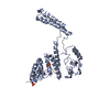
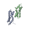
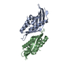
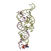
| Title | Structure of the CHIP-TPR domain in complex with the Hsc70 Lid-Tail domains | ||||||
 Components Components |
| ||||||
 Keywords Keywords | LIGASE/PROTEIN BINDING / TPR /  E3 ubiquitin ligase / Hsc70 / LIGASE-PROTEIN BINDING complex E3 ubiquitin ligase / Hsc70 / LIGASE-PROTEIN BINDING complex | ||||||
| Function / homology |  Function and homology information Function and homology informationpositive regulation of chaperone-mediated protein complex assembly / regulation of glucocorticoid metabolic process / lumenal side of lysosomal membrane / regulation of protein import / negative regulation of supramolecular fiber organization / positive regulation of protein refolding / prostaglandin binding / positive regulation of lysosomal membrane permeability / lysosomal matrix / A1 adenosine receptor binding ...positive regulation of chaperone-mediated protein complex assembly / regulation of glucocorticoid metabolic process / lumenal side of lysosomal membrane / regulation of protein import / negative regulation of supramolecular fiber organization / positive regulation of protein refolding / prostaglandin binding / positive regulation of lysosomal membrane permeability / lysosomal matrix / A1 adenosine receptor binding / protein carrier chaperone / Respiratory syncytial virus genome transcription / response to nickel cation / ubiquitin conjugating enzyme complex / clathrin-sculpted gamma-aminobutyric acid transport vesicle membrane / protein transmembrane import into intracellular organelle / Lipophagy / positive regulation of ERAD pathway / chaperone-mediated autophagy translocation complex disassembly / slow axonal transport / clathrin-uncoating ATPase activity / GABA synthesis, release, reuptake and degradation / protein targeting to lysosome involved in chaperone-mediated autophagy / late endosomal microautophagy / response to odorant / positive regulation of mitophagy / positive regulation by host of viral genome replication / synaptic vesicle uncoating / ERBB2 signaling pathway / C3HC4-type RING finger domain binding / positive regulation of smooth muscle cell apoptotic process / negative regulation of NLRP3 inflammasome complex assembly / clathrin coat disassembly / CHL1 interactions / ATP-dependent protein disaggregase activity / regulation of protein complex stability / nuclear inclusion body / photoreceptor ribbon synapse / maintenance of postsynaptic specialization structure / membrane organization / misfolded protein binding / Prp19 complex / glycinergic synapse / presynaptic cytosol / postsynaptic specialization membrane / protein folding chaperone complex / cellular response to misfolded protein / positive regulation of mRNA splicing, via spliceosome / ubiquitin-ubiquitin ligase activity / postsynaptic cytosol / regulation of postsynapse organization / RIPK1-mediated regulated necrosis / Lysosome Vesicle Biogenesis / negative regulation of cardiac muscle cell apoptotic process / intermediate filament / chaperone-mediated autophagy / cellular response to steroid hormone stimulus / Golgi Associated Vesicle Biogenesis / protein quality control for misfolded or incompletely synthesized proteins / TPR domain binding / phosphatidylserine binding / non-chaperonin molecular chaperone ATPase / ERAD pathway / SMAD binding / protein monoubiquitination / negative regulation of smooth muscle cell apoptotic process / chaperone cofactor-dependent protein refolding / R-SMAD binding / protein K63-linked ubiquitination / positive regulation of proteolysis / protein maturation / HSF1-dependent transactivation / response to unfolded protein / estrous cycle / Regulation of HSF1-mediated heat shock response / autophagosome / regulation of protein-containing complex assembly / Attenuation phase / protein autoubiquitination / Protein methylation / ATP metabolic process / protein folding chaperone / positive regulation of phagocytosis / ubiquitin ligase complex / endoplasmic reticulum unfolded protein response / skeletal muscle tissue development / forebrain development / Hsp70 protein binding / heat shock protein binding / HSP90 chaperone cycle for steroid hormone receptors (SHR) in the presence of ligand / cellular response to cadmium ion / cellular response to starvation / photoreceptor inner segment / lysosomal lumen / cerebellum development / Downregulation of TGF-beta receptor signaling / mRNA Splicing - Major Pathway / dendritic shaft / response to activity / positive regulation of protein ubiquitinationSimilarity search - Function | ||||||
| Biological species |  Homo sapiens (human) Homo sapiens (human) | ||||||
| Method | X-RAY DIFFRACTION / SYNCHROTRON / MOLECULAR REPLACEMENT / Resolution: 2.91 Å | ||||||
 Authors Authors | Page, R.C. / Amick, J. / Nix, J.C. / Misra, S. | ||||||
 Citation Citation | Journal: Structure / Year: 2015 Title: A Bipartite Interaction between Hsp70 and CHIP Regulates Ubiquitination of Chaperoned Client Proteins. Authors: Zhang, H. / Amick, J. / Chakravarti, R. / Santarriaga, S. / Schlanger, S. / McGlone, C. / Dare, M. / Nix, J.C. / Scaglione, K.M. / Stuehr, D.J. / Misra, S. / Page, R.C. | ||||||
| History |
|
- Structure visualization
| Structure viewer | Molecule: MolmilJmol/JSmol |
|---|
- Downloads & links
-Download
| PDBx/mmCIF format | 4kbq.cif.gz | 99.2 KB | Display | PDBx/mmCIF format |
|---|---|---|---|---|
| PDB format | pdb4kbq.ent.gz | 76.6 KB | Display | PDB format |
| PDBx/mmJSON format | 4kbq.json.gz | Tree view | PDBx/mmJSON format | |
| Others |  Other downloads Other downloads |
-Validation report
| Arichive directory | https://data.pdbj.org/pub/pdb/validation_reports/kb/4kbqftp://data.pdbj.org/pub/pdb/validation_reports/kb/4kbq | HTTPS FTP |
|---|
-Related structure data


-Links
 PDBj
PDBj


- Assembly
| Deposited unit | 
| ||||||||
|---|---|---|---|---|---|---|---|---|---|
| 1 | 
| ||||||||
| 2 | 
| ||||||||
| Unit cell |
|
-Components
| #1: Protein | Mass: 15884.057 Da / Num. of mol.: 2 / Fragment: TPR Source method: isolated from a genetically manipulated source Source: (gene. exp.) Homo sapiens (human) / Gene: CHIP, PP1131, STUB1 / Plasmid: pHis//2 / Production host:  Escherichia coli (E. coli) / Strain (production host): BL21(DE3) Rosetta Escherichia coli (E. coli) / Strain (production host): BL21(DE3) RosettaReferences: UniProt: Q9UNE7, Ligases; Forming carbon-nitrogen bonds; Acid-amino-acid ligases (peptide synthases)#2: Protein | Mass: 11423.743 Da / Num. of mol.: 2 / Fragment: Lid-Tail (delta626-638) / Mutation: delta(626-638) deletion mutant Source method: isolated from a genetically manipulated source Source: (gene. exp.) Homo sapiens (human) / Gene: HSC70, HSP73, HSPA10, HSPA8 / Plasmid: TOPO / Production host: Escherichia coli (E. coli) / Strain (production host): BL21(DE3) Rosetta / References: UniProt: P11142#3: Water | ChemComp-HOH / | Water Mass: 18.015 Da / Num. of mol.: 52 / Source method: isolated from a natural source / Formula: H2O Mass: 18.015 Da / Num. of mol.: 52 / Source method: isolated from a natural source / Formula: H2O |
|---|
-Experimental details
-Experiment
| Experiment | Method: X-RAY DIFFRACTION / Number of used crystals: 1 |
|---|
- Sample preparation
| Crystal | Density Matthews: 3.46 Å3/Da / Density % sol: 64.43 % |
|---|---|
| Crystal grow | Temperature: 293 K / Method: vapor diffusion, hanging drop / pH: 7 Details: 1.7M ammonium citrate, 0.1M HEPES, pH 7.0, VAPOR DIFFUSION, HANGING DROP, temperature 293K |
-Data collection
| Diffraction | Mean temperature: 100 K |
|---|---|
| Diffraction source | Source: SYNCHROTRON / Site: ALS  / Beamline: 4.2.2 / Wavelength: 1 / Beamline: 4.2.2 / Wavelength: 1 |
| Detector | Type: NOIR-1 / Detector: CCD / Date: Oct 17, 2012 |
| Radiation | Monochromator: Rosenbaum-Rock Si(111) sagitally focused monochromator Protocol: SINGLE WAVELENGTH / Monochromatic (M) / Laue (L): M / Scattering type: x-ray |
| Radiation wavelength | Wavelength: 1 Å / Relative weight: 1 |
| Reflection | Resolution: 2.91→64.75 Å / Num. obs: 17188 / % possible obs: 97.8 % / Observed criterion σ(F): 2 / Observed criterion σ(I): 2 |
| Reflection shell | Resolution: 2.91→3.01 Å / Redundancy: 12.1 % / Rmerge(I) obs: 0.639 / Mean I/σ(I) obs: 4.2 / % possible all: 76.7 |
- Processing
| Software |
| |||||||||||||||||||||||||||||||||||||||||||||||||||||||||||||||||||||||||||||||||||||||||||||||||||||||||||||||||||||||||||||||||||||||||||||||||||||||||||||||||
|---|---|---|---|---|---|---|---|---|---|---|---|---|---|---|---|---|---|---|---|---|---|---|---|---|---|---|---|---|---|---|---|---|---|---|---|---|---|---|---|---|---|---|---|---|---|---|---|---|---|---|---|---|---|---|---|---|---|---|---|---|---|---|---|---|---|---|---|---|---|---|---|---|---|---|---|---|---|---|---|---|---|---|---|---|---|---|---|---|---|---|---|---|---|---|---|---|---|---|---|---|---|---|---|---|---|---|---|---|---|---|---|---|---|---|---|---|---|---|---|---|---|---|---|---|---|---|---|---|---|---|---|---|---|---|---|---|---|---|---|---|---|---|---|---|---|---|---|---|---|---|---|---|---|---|---|---|---|---|---|---|---|---|
| Refinement | Method to determine structure: MOLECULAR REPLACEMENT Starting model: PDB ENTRIES 2C2L, 3LOF Resolution: 2.91→64.746 Å / SU ML: 0.41 / σ(F): 1.6 / Phase error: 24.54 / Stereochemistry target values: ML
| |||||||||||||||||||||||||||||||||||||||||||||||||||||||||||||||||||||||||||||||||||||||||||||||||||||||||||||||||||||||||||||||||||||||||||||||||||||||||||||||||
| Solvent computation | Shrinkage radii: 0.9 Å / VDW probe radii: 1.11 Å / Solvent model: FLAT BULK SOLVENT MODEL | |||||||||||||||||||||||||||||||||||||||||||||||||||||||||||||||||||||||||||||||||||||||||||||||||||||||||||||||||||||||||||||||||||||||||||||||||||||||||||||||||
| Refinement step | Cycle: LAST / Resolution: 2.91→64.746 Å
| |||||||||||||||||||||||||||||||||||||||||||||||||||||||||||||||||||||||||||||||||||||||||||||||||||||||||||||||||||||||||||||||||||||||||||||||||||||||||||||||||
| Refine LS restraints |
| |||||||||||||||||||||||||||||||||||||||||||||||||||||||||||||||||||||||||||||||||||||||||||||||||||||||||||||||||||||||||||||||||||||||||||||||||||||||||||||||||
| LS refinement shell |
|
