 Movie
Movie Controller

[English] 日本語
 Yorodumi
- PDB-4kbm: Structure of the Mtb CarD/RNAP Beta subunit B1-B2 domains complex -
+ Open data

- Basic information
| Entry | Database: PDB / ID: 4kbm | ||||||
|---|---|---|---|---|---|---|---|
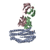
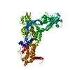
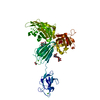
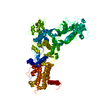
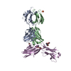
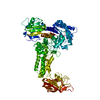
| Title | Structure of the Mtb CarD/RNAP Beta subunit B1-B2 domains complex | ||||||
 Components Components |
| ||||||
 Keywords Keywords | TRANSFERASE/TRANSCRIPTION /  Structural Genomics / TB Structural Genomics Consortium / TBSGC / Tudor / DNA-dependent RNA polymerase / transcription regulator / protein-protein complex / CarD / DNA / sigma factor / TRANSFERASE-TRANSCRIPTION complex Structural Genomics / TB Structural Genomics Consortium / TBSGC / Tudor / DNA-dependent RNA polymerase / transcription regulator / protein-protein complex / CarD / DNA / sigma factor / TRANSFERASE-TRANSCRIPTION complex | ||||||
| Function / homology |  Function and homology information Function and homology informationregulation of growth rate / Antimicrobial action and antimicrobial resistance in Mtb / cell wall / stringent response / rRNA transcription / DNA-directed RNA polymerase complex / peptidoglycan-based cell wall / ribonucleoside binding / DNA-directed 5'-3' RNA polymerase activity / DNA-directed RNA polymerase ...regulation of growth rate / Antimicrobial action and antimicrobial resistance in Mtb / cell wall / stringent response / rRNA transcription / DNA-directed RNA polymerase complex / peptidoglycan-based cell wall / ribonucleoside binding / DNA-directed 5'-3' RNA polymerase activity / DNA-directed RNA polymerase / response to antibiotic / DNA-templated transcription / DNA binding / plasma membrane / cytosolSimilarity search - Function | ||||||
| Biological species |   Mycobacterium tuberculosis (bacteria) Mycobacterium tuberculosis (bacteria) | ||||||
| Method | X-RAY DIFFRACTION / SYNCHROTRON / MOLECULAR REPLACEMENT / Resolution: 2.1146 Å | ||||||
 Authors Authors | Gulten, G. / Sacchettini, J.C. / TB Structural Genomics Consortium (TBSGC) | ||||||
 Citation Citation | Journal: Structure / Year: 2013 Title: Structure of the Mtb CarD/RNAP beta-Lobes Complex Reveals the Molecular Basis of Interaction and Presents a Distinct DNA-Binding Domain for Mtb CarD. Authors: Gulten, G. / Sacchettini, J.C. | ||||||
| History |
|
- Structure visualization
| Structure viewer | Molecule: MolmilJmol/JSmol |
|---|
- Downloads & links
-Download
| PDBx/mmCIF format | 4kbm.cif.gz | 217.8 KB | Display | PDBx/mmCIF format |
|---|---|---|---|---|
| PDB format | pdb4kbm.ent.gz | 173.4 KB | Display | PDB format |
| PDBx/mmJSON format | 4kbm.json.gz | Tree view | PDBx/mmJSON format | |
| Others |  Other downloads Other downloads |
-Validation report
| Arichive directory | https://data.pdbj.org/pub/pdb/validation_reports/kb/4kbmftp://data.pdbj.org/pub/pdb/validation_reports/kb/4kbm | HTTPS FTP |
|---|
-Related structure data
| Related structure data | 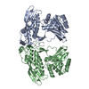 4kbjSC S: Starting model for refinement C: citing same article ( |
|---|---|
| Similar structure data |


-Links
 PDBj
PDBj


- Assembly
| Deposited unit | 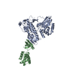
| ||||||||
|---|---|---|---|---|---|---|---|---|---|
| 1 |
| ||||||||
| Unit cell |
|
-Components
| #1: Protein | Polymerase / RNAP subunit beta / RNA polymerase subunit beta / Transcriptase subunit beta Mass: 46266.324 Da / Num. of mol.: 1 / Fragment: B1 and B2 domains of Mtb RNAP Source method: isolated from a genetically manipulated source Source: (gene. exp.) Mycobacterium tuberculosis (bacteria) / Strain: H37Rv / Gene: MT0695, MTCI376.08c, rpoB, Rv0667 / Plasmid: pET15b / Production host: Escherichia coli (E. coli) / Strain (production host): Rosetta2(DE3)pLysSReferences: UniProt: P0A680, UniProt: P9WGY9*PLUS, DNA-directed RNA polymerase |
|---|---|
| #2: Protein | Mass: 17933.361 Da / Num. of mol.: 1 Source method: isolated from a genetically manipulated source Source: (gene. exp.) Mycobacterium tuberculosis (bacteria) / Strain: H37Rv / Gene: carD, MT3689, Rv3583c / Plasmid: pET30b / Production host: Escherichia coli (E. coli) / Strain (production host): Rosetta2(DE3)pLysS / References: UniProt: O53568, UniProt: P9WJG3*PLUS |
| #3: Water | ChemComp-HOH / Water Mass: 18.015 Da / Num. of mol.: 305 / Source method: isolated from a natural source / Formula: H2O Mass: 18.015 Da / Num. of mol.: 305 / Source method: isolated from a natural source / Formula: H2O |
-Experimental details
-Experiment
| Experiment | Method: X-RAY DIFFRACTION / Number of used crystals: 1 |
|---|
- Sample preparation
| Crystal | Density Matthews: 2.78 Å3/Da / Density % sol: 55.78 % |
|---|---|
| Crystal grow | Temperature: 291 K / Method: vapor diffusion, hanging drop / pH: 5.6 Details: 14% PEG 3350, 2% tacsimate pH 5.0, 0.1 M sodium citrate tribasic dihydrate, VAPOR DIFFUSION, HANGING DROP, temperature 291K |
-Data collection
| Diffraction | Mean temperature: 100 K |
|---|---|
| Diffraction source | Source: SYNCHROTRON / Site: APS  / Beamline: 23-ID-D / Wavelength: 0.979 Å / Beamline: 23-ID-D / Wavelength: 0.979 Å |
| Detector | Type: MAR 300 / Detector: CCD / Date: Jun 9, 2012 |
| Radiation | Monochromator: Si(111) / Protocol: SINGLE WAVELENGTH / Monochromatic (M) / Laue (L): M / Scattering type: x-ray |
| Radiation wavelength | Wavelength: 0.979 Å / Relative weight: 1 |
| Reflection | Resolution: 2.11→37.59 Å / Num. all: 40844 / Num. obs: 40844 / % possible obs: 98.1 % / Observed criterion σ(F): 2 / Observed criterion σ(I): 2 |
| Reflection shell | Resolution: 2.11→2.21 Å / Mean I/σ(I) obs: 2.03 / Num. unique all: 4675 / % possible all: 92 |
- Processing
| Software |
| ||||||||||||||||||||||||||||||||||||||||||||||||||||||||||||||||||||||||||||||||||||||||||||||||||||||||||||||||||||||||||||||||||||||||||||||||||||||
|---|---|---|---|---|---|---|---|---|---|---|---|---|---|---|---|---|---|---|---|---|---|---|---|---|---|---|---|---|---|---|---|---|---|---|---|---|---|---|---|---|---|---|---|---|---|---|---|---|---|---|---|---|---|---|---|---|---|---|---|---|---|---|---|---|---|---|---|---|---|---|---|---|---|---|---|---|---|---|---|---|---|---|---|---|---|---|---|---|---|---|---|---|---|---|---|---|---|---|---|---|---|---|---|---|---|---|---|---|---|---|---|---|---|---|---|---|---|---|---|---|---|---|---|---|---|---|---|---|---|---|---|---|---|---|---|---|---|---|---|---|---|---|---|---|---|---|---|---|---|---|---|
| Refinement | Method to determine structure: MOLECULAR REPLACEMENT Starting model: PDB ENTRY 4KBJ Resolution: 2.1146→37.588 Å / SU ML: 0.22 / σ(F): 1.34 / Phase error: 23.48 / Stereochemistry target values: ML
| ||||||||||||||||||||||||||||||||||||||||||||||||||||||||||||||||||||||||||||||||||||||||||||||||||||||||||||||||||||||||||||||||||||||||||||||||||||||
| Solvent computation | Shrinkage radii: 0.86 Å / VDW probe radii: 1.1 Å / Solvent model: FLAT BULK SOLVENT MODEL / Bsol: 33.559 Å2 / ksol: 0.321 e/Å3 | ||||||||||||||||||||||||||||||||||||||||||||||||||||||||||||||||||||||||||||||||||||||||||||||||||||||||||||||||||||||||||||||||||||||||||||||||||||||
| Displacement parameters |
| ||||||||||||||||||||||||||||||||||||||||||||||||||||||||||||||||||||||||||||||||||||||||||||||||||||||||||||||||||||||||||||||||||||||||||||||||||||||
| Refinement step | Cycle: LAST / Resolution: 2.1146→37.588 Å
| ||||||||||||||||||||||||||||||||||||||||||||||||||||||||||||||||||||||||||||||||||||||||||||||||||||||||||||||||||||||||||||||||||||||||||||||||||||||
| Refine LS restraints |
| ||||||||||||||||||||||||||||||||||||||||||||||||||||||||||||||||||||||||||||||||||||||||||||||||||||||||||||||||||||||||||||||||||||||||||||||||||||||
| LS refinement shell |
| ||||||||||||||||||||||||||||||||||||||||||||||||||||||||||||||||||||||||||||||||||||||||||||||||||||||||||||||||||||||||||||||||||||||||||||||||||||||
| Refinement TLS params. | Method: refined / Refine-ID: X-RAY DIFFRACTION
| ||||||||||||||||||||||||||||||||||||||||||||||||||||||||||||||||||||||||||||||||||||||||||||||||||||||||||||||||||||||||||||||||||||||||||||||||||||||
| Refinement TLS group |
|
