 Movie
Movie Controller

[English] 日本語
 Yorodumi
- PDB-4zso: Crystal structure of a complex between B7-H6, a tumor cell ligand... -
+ Open data

- Basic information
| Entry | Database: PDB / ID: 4zso | |||||||||
|---|---|---|---|---|---|---|---|---|---|---|
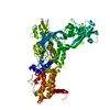
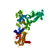
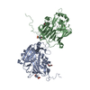
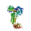
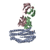
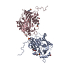
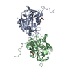
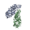
| Title | Crystal structure of a complex between B7-H6, a tumor cell ligand for natural cytotoxicity receptor NKp30, and an inhibitory antibody | |||||||||
 Components Components |
| |||||||||
 Keywords Keywords |  IMMUNE SYSTEM / Tumor ligand B7-H6 / Antibody IMMUNE SYSTEM / Tumor ligand B7-H6 / Antibody | |||||||||
| Function / homology |  Function and homology information Function and homology informationimmunoglobulin complex, circulating / immunoglobulin receptor binding / complement activation, classical pathway / antigen binding / Immunoregulatory interactions between a Lymphoid and a non-Lymphoid cell / antibacterial humoral response / plasma membraneSimilarity search - Function | |||||||||
| Biological species |  Homo sapiens (human) Homo sapiens (human) | |||||||||
| Method | X-RAY DIFFRACTION / SYNCHROTRON / MOLECULAR REPLACEMENT / Resolution: 2.5 Å | |||||||||
 Authors Authors | Xu, X. / Li, Y. / Mariuzza, R.A. | |||||||||
 Citation Citation | Journal: to be published Title: Crystal structure of a complex between B7-H6, a tumor cell ligand for natural cytotoxicity receptor NKp30, and an inhibitory antibody Authors: Xu, X. / Li, Y. / Vivier, E. / Chen, Q. / Mariuzza, R.A. | |||||||||
| History |
|
- Structure visualization
| Structure viewer | Molecule: MolmilJmol/JSmol |
|---|
- Downloads & links
-Download
| PDBx/mmCIF format | 4zso.cif.gz | 260.4 KB | Display | PDBx/mmCIF format |
|---|---|---|---|---|
| PDB format | pdb4zso.ent.gz | 211.8 KB | Display | PDB format |
| PDBx/mmJSON format | 4zso.json.gz | Tree view | PDBx/mmJSON format | |
| Others |  Other downloads Other downloads |
-Validation report
| Arichive directory | https://data.pdbj.org/pub/pdb/validation_reports/zs/4zsoftp://data.pdbj.org/pub/pdb/validation_reports/zs/4zso | HTTPS FTP |
|---|
-Related structure data
| Similar structure data |
|---|


-Links
 PDBj
PDBj


- Assembly
| Deposited unit | 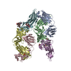
| ||||||||
|---|---|---|---|---|---|---|---|---|---|
| 1 | 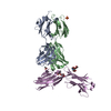
| ||||||||
| 2 | 
| ||||||||
| Unit cell |
|
-Components
-Protein , 1 types, 2 molecules EF
| #3: Protein | Mass: 28319.947 Da / Num. of mol.: 2 Source method: isolated from a genetically manipulated source Source: (gene. exp.) Homo sapiens (human) / Gene: NCR3LG1, B7H6 / Production host:  unidentified baculovirus / References: UniProt: Q68D85 unidentified baculovirus / References: UniProt: Q68D85 |
|---|
-Antibody , 2 types, 4 molecules ACBD
| #1: Antibody | Immunoglobulin light chain Mass: 23508.086 Da / Num. of mol.: 2 Source method: isolated from a genetically manipulated source Source: (gene. exp.) Homo sapiens (human) / Plasmid: pET-26b / Production host:  Escherichia Coli (E. coli) / Strain (production host): BL21(DE3) Escherichia Coli (E. coli) / Strain (production host): BL21(DE3)#2: Antibody | Immunoglobulin heavy chainMass: 23249.070 Da / Num. of mol.: 2 Source method: isolated from a genetically manipulated source Source: (gene. exp.) Homo sapiens (human) / Plasmid: pET-26b / Production host: Escherichia coli (E. coli) / Strain (production host): BL21(DE3) |
|---|
-Sugars , 3 types, 4 molecules
| #4: Polysaccharide | alpha-L-fucopyranose-(1-6)-2-acetamido-2-deoxy-beta-D-glucopyranose / Mass: 367.349 Da / Num. of mol.: 1 Source method: isolated from a genetically manipulated source | ||
|---|---|---|---|
| #5: Polysaccharide | / Mass: 424.401 Da / Num. of mol.: 2 Source method: isolated from a genetically manipulated source #6: Polysaccharide | 2-acetamido-2-deoxy-beta-D-glucopyranose-(1-4)-[alpha-L-fucopyranose-(1-6)]2-acetamido-2-deoxy-beta- ...2-acetamido-2-deoxy-beta-D-glucopyranose-(1-4)-[alpha-L-fucopyranose-(1-6)]2-acetamido-2-deoxy-beta-D-glucopyranose | / Mass: 570.542 Da / Num. of mol.: 1Source method: isolated from a genetically manipulated source |
-Non-polymers , 3 types, 255 molecules 


| #7: Chemical | Sulfate Mass: 96.063 Da / Num. of mol.: 2 / Source method: obtained synthetically / Formula: SO4 Mass: 96.063 Da / Num. of mol.: 2 / Source method: obtained synthetically / Formula: SO4#8: Chemical | Acetic acid Mass: 60.052 Da / Num. of mol.: 2 / Source method: obtained synthetically / Formula: C2H4O2 Mass: 60.052 Da / Num. of mol.: 2 / Source method: obtained synthetically / Formula: C2H4O2#9: Water | ChemComp-HOH / | WaterMass: 18.015 Da / Num. of mol.: 251 / Source method: isolated from a natural source / Formula: H2O |
|---|
-Experimental details
-Experiment
| Experiment | Method: X-RAY DIFFRACTION / Number of used crystals: 1 |
|---|
- Sample preparation
| Crystal | Density Matthews: 3.53 Å3/Da / Density % sol: 65.17 % |
|---|---|
| Crystal grow | Temperature: 298 K / Method: evaporation / pH: 8 Details: PEG 8000, Tris-HCl, lithium sulfate, calcium acetate PH range: 7.0-8.5 |
-Data collection
| Diffraction | Mean temperature: 100 K |
|---|---|
| Diffraction source | Source: SYNCHROTRON / Site: APS  / Beamline: 19-ID / Wavelength: 1.502 Å / Beamline: 19-ID / Wavelength: 1.502 Å |
| Detector | Type: ADSC QUANTUM 4 / Detector: CCD / Date: Aug 20, 2014 / Details: mirrors |
| Radiation | Monochromator: GRAPHITE / Protocol: SINGLE WAVELENGTH / Monochromatic (M) / Laue (L): M / Scattering type: x-ray |
| Radiation wavelength | Wavelength: 1.502 Å / Relative weight: 1 |
| Reflection | Resolution: 2.5→50 Å / Num. obs: 74515 / % possible obs: 100 % / Redundancy: 14.4 % / Rmerge(I) obs: 0.143 / Net I/σ(I): 6.3 |
| Reflection shell | Resolution: 2.5→2.59 Å / Redundancy: 13.3 % / Rmerge(I) obs: 0.783 / % possible all: 99.8 |
- Processing
| Software |
| ||||||||||||||||||||||||||||||||||||||||||||||||||||||||||||
|---|---|---|---|---|---|---|---|---|---|---|---|---|---|---|---|---|---|---|---|---|---|---|---|---|---|---|---|---|---|---|---|---|---|---|---|---|---|---|---|---|---|---|---|---|---|---|---|---|---|---|---|---|---|---|---|---|---|---|---|---|---|
| Refinement | Method to determine structure: MOLECULAR REPLACEMENT / Resolution: 2.5→50 Å / Cross valid method: FREE R-VALUE / σ(F): 0
| ||||||||||||||||||||||||||||||||||||||||||||||||||||||||||||
| Solvent computation | Bsol: 32.23 Å2 | ||||||||||||||||||||||||||||||||||||||||||||||||||||||||||||
| Displacement parameters | Biso mean: 43.45 Å2
| ||||||||||||||||||||||||||||||||||||||||||||||||||||||||||||
| Refinement step | Cycle: LAST / Resolution: 2.5→50 Å
| ||||||||||||||||||||||||||||||||||||||||||||||||||||||||||||
| Refine LS restraints |
| ||||||||||||||||||||||||||||||||||||||||||||||||||||||||||||
| Xplor file |
|
