 Movie
Movie Controller


+ Open data

- Basic information
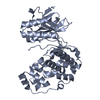
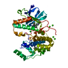
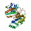
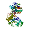
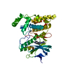
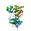
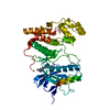
| Entry | Database: PDB / ID: 4ka3 | ||||||
|---|---|---|---|---|---|---|---|
| Title | Structure of MAP kinase in complex with a docking peptide | ||||||
 Components Components |
| ||||||
 Keywords Keywords | TRANSFERASE/PROTEIN BINDING /  kinase domain / phosphorylation / KIM / TRANSFERASE-PROTEIN BINDING complex kinase domain / phosphorylation / KIM / TRANSFERASE-PROTEIN BINDING complex | ||||||
| Function / homology |  Function and homology information Function and homology informationcardiac septum development / DSCAM interactions / p38MAPK events / Activation of the AP-1 family of transcription factors / Platelet sensitization by LDL / RHO GTPases Activate NADPH Oxidases / ERK/MAPK targets / activated TAK1 mediates p38 MAPK activation / NOD1/2 Signaling Pathway / ADP signalling through P2Y purinoceptor 1 ...cardiac septum development / DSCAM interactions / p38MAPK events / Activation of the AP-1 family of transcription factors / Platelet sensitization by LDL / RHO GTPases Activate NADPH Oxidases / ERK/MAPK targets / activated TAK1 mediates p38 MAPK activation / NOD1/2 Signaling Pathway / ADP signalling through P2Y purinoceptor 1 / Oxidative Stress Induced Senescence / Regulation of TP53 Activity through Phosphorylation / Myogenesis / coronary vasculature development / VEGFA-VEGFR2 Pathway / aorta development / regulation of synaptic membrane adhesion / stress-induced premature senescence / cell surface receptor protein serine/threonine kinase signaling pathway / cartilage condensation / cellular response to UV-B / protein serine/threonine phosphatase activity / mitogen-activated protein kinase p38 binding / positive regulation of myoblast fusion / negative regulation of hippo signaling / positive regulation of myotube differentiation / NFAT protein binding / glucose import / regulation of cytokine production involved in inflammatory response / p38MAPK cascade / non-canonical NF-kappaB signal transduction / fatty acid oxidation / cellular response to lipoteichoic acid / response to dietary excess / response to muramyl dipeptide / MAP kinase activity / regulation of ossification / mitogen-activated protein kinase / cellular response to vascular endothelial growth factor stimulus / positive regulation of myoblast differentiation / chondrocyte differentiation / vascular endothelial growth factor receptor signaling pathway / enzyme activator activity / stress-activated MAPK cascade / heart morphogenesis / skeletal muscle tissue development / lipopolysaccharide-mediated signaling pathway / positive regulation of cardiac muscle cell proliferation / IRAK2 mediated activation of TAK1 complex / Alpha-protein kinase 1 signaling pathway / IRAK2 mediated activation of TAK1 complex upon TLR7/8 or 9 stimulation / TICAM1,TRAF6-dependent induction of TAK1 complex / striated muscle cell differentiation / response to muscle stretch / TRAF6-mediated induction of TAK1 complex within TLR4 complex / positive regulation of brown fat cell differentiation / Neutrophil degranulation / positive regulation of interleukin-12 production / protein serine/threonine kinase activator activity / transforming growth factor beta receptor signaling pathway / osteoclast differentiation / JNK (c-Jun kinases) phosphorylation and activation mediated by activated human TAK1 / positive regulation of erythrocyte differentiation / TNFR1-induced NF-kappa-B signaling pathway / DNA damage checkpoint signaling / activated TAK1 mediates p38 MAPK activation / cellular response to ionizing radiation / stem cell differentiation / positive regulation of glucose import / placenta development / TAK1-dependent IKK and NF-kappa-B activation / lung development / NOD1/2 Signaling Pathway / response to insulin / bone development / positive regulation of protein serine/threonine kinase activity / negative regulation of canonical Wnt signaling pathway / cell morphogenesis / CLEC7A (Dectin-1) signaling / cellular response to virus / FCERI mediated NF-kB activation / spindle pole / positive regulation of protein import into nucleus / Interleukin-1 signaling / osteoblast differentiation / glucose metabolic process / positive regulation of reactive oxygen species metabolic process / MAPK cascade / cellular response to tumor necrosis factor / kinase activity / peptidyl-serine phosphorylation / protein phosphatase binding / angiogenesis / in utero embryonic development / cellular response to lipopolysaccharide / response to lipopolysaccharide / transcription by RNA polymerase II / positive regulation of MAPK cascade / molecular adaptor activity / endosome membraneSimilarity search - Function | ||||||
| Biological species |  Mus musculus (house mouse) Mus musculus (house mouse) Homo sapiens (human) Homo sapiens (human) | ||||||
| Method | X-RAY DIFFRACTION / SYNCHROTRON / MOLECULAR REPLACEMENT / Resolution: 2.707 Å | ||||||
 Authors Authors | Xin, F.J. / Wu, J.W. | ||||||
 Citation Citation | Journal: Sci China Life Sci / Year: 2013 Title: Crystal structure of the p38 alpha MAP kinase in complex with a docking peptide from TAB1 Authors: Xin, F.J. / Wu, J.W. | ||||||
| History |
|
- Structure visualization
| Structure viewer | Molecule: MolmilJmol/JSmol |
|---|
- Downloads & links
-Download
| PDBx/mmCIF format | 4ka3.cif.gz | 152.9 KB | Display | PDBx/mmCIF format |
|---|---|---|---|---|
| PDB format | pdb4ka3.ent.gz | 122.1 KB | Display | PDB format |
| PDBx/mmJSON format | 4ka3.json.gz | Tree view | PDBx/mmJSON format | |
| Others |  Other downloads Other downloads |
-Validation report
| Arichive directory | https://data.pdbj.org/pub/pdb/validation_reports/ka/4ka3ftp://data.pdbj.org/pub/pdb/validation_reports/ka/4ka3 | HTTPS FTP |
|---|
-Related structure data
| Related structure data |  1lezS S: Starting model for refinement |
|---|---|
| Similar structure data |


-Links
 PDBj
PDBj


- Assembly
| Deposited unit | 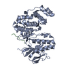
| ||||||||
|---|---|---|---|---|---|---|---|---|---|
| 1 |
| ||||||||
| Unit cell |
|
-Components
| #1: Protein | MAPK14 / MAP kinase 14 / MAPK 14 / CRK1 / Mitogen-activated protein kinase p38 alpha / MAP kinase p38 alpha Mass: 41338.160 Da / Num. of mol.: 1 Source method: isolated from a genetically manipulated source Source: (gene. exp.) Mus musculus (house mouse) / Gene: Mapk14 / Plasmid: pET21b / Production host:  Escherichia coli (E. coli) / Strain (production host): BL21(DE3) Escherichia coli (E. coli) / Strain (production host): BL21(DE3)References: UniProt: P47811, mitogen-activated protein kinase |
|---|---|
| #2: Protein/peptide | Mass: 3168.479 Da / Num. of mol.: 1 / Fragment: UNP residues 395-415 Source method: isolated from a genetically manipulated source Source: (gene. exp.) Homo sapiens (human) / Gene: TAB1 / Plasmid: pET21b / Production host: Escherichia coli (E. coli) / Strain (production host): BL21(DE3) / References: UniProt: Q15750 |
| #3: Water | ChemComp-HOH / Water Mass: 18.015 Da / Num. of mol.: 8 / Source method: isolated from a natural source / Formula: H2O Mass: 18.015 Da / Num. of mol.: 8 / Source method: isolated from a natural source / Formula: H2O |
-Experimental details
-Experiment
| Experiment | Method: X-RAY DIFFRACTION / Number of used crystals: 1 |
|---|
- Sample preparation
| Crystal | Density Matthews: 2.68 Å3/Da / Density % sol: 54.14 % |
|---|---|
| Crystal grow | Temperature: 294 K / Method: vapor diffusion, hanging drop / pH: 7.86 Details: 100mM Hepes, 22% polyacrylic acid 5100, pH 7.86, VAPOR DIFFUSION, HANGING DROP, temperature 294K |
-Data collection
| Diffraction | Mean temperature: 100 K |
|---|---|
| Diffraction source | Source: SYNCHROTRON / Site: SSRF  / Beamline: BL17U / Wavelength: 0.979413 Å / Beamline: BL17U / Wavelength: 0.979413 Å |
| Detector | Type: ADSC QUANTUM 315 / Detector: CCD / Date: Mar 28, 2011 |
| Radiation | Monochromator: GRAPHITE / Protocol: SINGLE WAVELENGTH / Monochromatic (M) / Laue (L): M / Scattering type: x-ray |
| Radiation wavelength | Wavelength: 0.979413 Å / Relative weight: 1 |
| Reflection | Resolution: 2.707→50 Å / Num. all: 13030 / Num. obs: 13001 / % possible obs: 96.1 % / Observed criterion σ(F): 1 / Observed criterion σ(I): 1 / Redundancy: 5.5 % / Biso Wilson estimate: 61.93 Å2 / Rmerge(I) obs: 0.061 / Net I/σ(I): 21.1 |
| Reflection shell | Resolution: 2.71→2.76 Å / Redundancy: 6 % / Rmerge(I) obs: 0.457 / Mean I/σ(I) obs: 4.9 / Num. unique all: 662 / % possible all: 100 |
- Processing
| Software |
| ||||||||||||||||||||||||||||||||||||||||||||||||||||||||||||||||||||||||||||||||||||||||||||||||||||||||||||||||||||||||||||||||||||||||||||||||||||||
|---|---|---|---|---|---|---|---|---|---|---|---|---|---|---|---|---|---|---|---|---|---|---|---|---|---|---|---|---|---|---|---|---|---|---|---|---|---|---|---|---|---|---|---|---|---|---|---|---|---|---|---|---|---|---|---|---|---|---|---|---|---|---|---|---|---|---|---|---|---|---|---|---|---|---|---|---|---|---|---|---|---|---|---|---|---|---|---|---|---|---|---|---|---|---|---|---|---|---|---|---|---|---|---|---|---|---|---|---|---|---|---|---|---|---|---|---|---|---|---|---|---|---|---|---|---|---|---|---|---|---|---|---|---|---|---|---|---|---|---|---|---|---|---|---|---|---|---|---|---|---|---|
| Refinement | Method to determine structure: MOLECULAR REPLACEMENT Starting model: 1LEZ Resolution: 2.707→26.827 Å / Occupancy max: 1 / Occupancy min: 1 / FOM work R set: 0.79 / SU ML: 0.44 / σ(F): 1.34 / Phase error: 26.99 / Stereochemistry target values: ML
| ||||||||||||||||||||||||||||||||||||||||||||||||||||||||||||||||||||||||||||||||||||||||||||||||||||||||||||||||||||||||||||||||||||||||||||||||||||||
| Solvent computation | Shrinkage radii: 0.73 Å / VDW probe radii: 1 Å / Solvent model: FLAT BULK SOLVENT MODEL / Bsol: 34.625 Å2 / ksol: 0.313 e/Å3 | ||||||||||||||||||||||||||||||||||||||||||||||||||||||||||||||||||||||||||||||||||||||||||||||||||||||||||||||||||||||||||||||||||||||||||||||||||||||
| Displacement parameters | Biso max: 121.07 Å2 / Biso mean: 70.358 Å2 / Biso min: 30 Å2
| ||||||||||||||||||||||||||||||||||||||||||||||||||||||||||||||||||||||||||||||||||||||||||||||||||||||||||||||||||||||||||||||||||||||||||||||||||||||
| Refinement step | Cycle: LAST / Resolution: 2.707→26.827 Å
| ||||||||||||||||||||||||||||||||||||||||||||||||||||||||||||||||||||||||||||||||||||||||||||||||||||||||||||||||||||||||||||||||||||||||||||||||||||||
| Refine LS restraints |
| ||||||||||||||||||||||||||||||||||||||||||||||||||||||||||||||||||||||||||||||||||||||||||||||||||||||||||||||||||||||||||||||||||||||||||||||||||||||
| LS refinement shell | Refine-ID: X-RAY DIFFRACTION / Total num. of bins used: 5
| ||||||||||||||||||||||||||||||||||||||||||||||||||||||||||||||||||||||||||||||||||||||||||||||||||||||||||||||||||||||||||||||||||||||||||||||||||||||
| Refinement TLS params. | Method: refined / Refine-ID: X-RAY DIFFRACTION
| ||||||||||||||||||||||||||||||||||||||||||||||||||||||||||||||||||||||||||||||||||||||||||||||||||||||||||||||||||||||||||||||||||||||||||||||||||||||
| Refinement TLS group |
|
