 Movie
Movie Controller


+ Open data

- Basic information
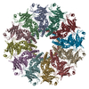
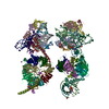
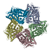
| Entry | Database: PDB / ID: 4k7h | ||||||
|---|---|---|---|---|---|---|---|
| Title | Major capsid protein P1 of the Pseudomonas phage phi6 | ||||||
 Components Components | Major inner protein P1 | ||||||
 Keywords Keywords |  VIRAL PROTEIN / major capsid protein VIRAL PROTEIN / major capsid protein | ||||||
| Function / homology | : / Major inner capsid protein P1 / T=2 icosahedral viral capsid / viral inner capsid / viral nucleocapsid / RNA binding / identical protein binding / Major inner protein P1 Function and homology information Function and homology information | ||||||
| Biological species |  Pseudomonas phage phi6 (bacteriophage) Pseudomonas phage phi6 (bacteriophage) | ||||||
| Method | X-RAY DIFFRACTION / SYNCHROTRON / MOLECULAR REPLACEMENT / Resolution: 3.5964 Å | ||||||
 Authors Authors | Boura, E. / Nemecek, D. / Plevka, P. / Steven, C.A. / Hurley, J.H. | ||||||
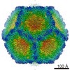
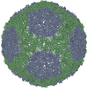
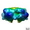
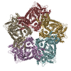
 Citation Citation | Journal: Structure / Year: 2013 Title: Subunit folds and maturation pathway of a dsRNA virus capsid. Authors: Daniel Nemecek / Evzen Boura / Weimin Wu / Naiqian Cheng / Pavel Plevka / Jian Qiao / Leonard Mindich / J Bernard Heymann / James H Hurley / Alasdair C Steven /  Abstract: The cystovirus ϕ6 shares several distinct features with other double-stranded RNA (dsRNA) viruses, including the human pathogen, rotavirus: segmented genomes, nonequivalent packing of 120 subunits ...The cystovirus ϕ6 shares several distinct features with other double-stranded RNA (dsRNA) viruses, including the human pathogen, rotavirus: segmented genomes, nonequivalent packing of 120 subunits in its icosahedral capsid, and capsids as compartments for transcription and replication. ϕ6 assembles as a dodecahedral procapsid that undergoes major conformational changes as it matures into the spherical capsid. We determined the crystal structure of the capsid protein, P1, revealing a flattened trapezoid subunit with an α-helical fold. We also solved the procapsid with cryo-electron microscopy to comparable resolution. Fitting the crystal structure into the procapsid disclosed substantial conformational differences between the two P1 conformers. Maturation via two intermediate states involves remodeling on a similar scale, besides huge rigid-body rotations. The capsid structure and its stepwise maturation that is coupled to sequential packaging of three RNA segments sets the cystoviruses apart from other dsRNA viruses as a dynamic molecular machine. | ||||||
| History |
|
- Structure visualization
| Structure viewer | Molecule: MolmilJmol/JSmol |
|---|
- Downloads & links
-Download
| PDBx/mmCIF format | 4k7h.cif.gz | 711.8 KB | Display | PDBx/mmCIF format |
|---|---|---|---|---|
| PDB format | pdb4k7h.ent.gz | 596.6 KB | Display | PDB format |
| PDBx/mmJSON format | 4k7h.json.gz | Tree view | PDBx/mmJSON format | |
| Others |  Other downloads Other downloads |
-Validation report
| Arichive directory | https://data.pdbj.org/pub/pdb/validation_reports/k7/4k7hftp://data.pdbj.org/pub/pdb/validation_reports/k7/4k7h | HTTPS FTP |
|---|
-Related structure data


-Links
 PDBj
PDBj
- Assembly
| Deposited unit | 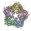
| ||||||||
|---|---|---|---|---|---|---|---|---|---|
| 1 |
| ||||||||
| 2 | 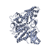
| ||||||||
| 3 | 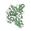
| ||||||||
| 4 | 
| ||||||||
| 5 | 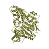
| ||||||||
| 6 | 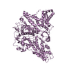
| ||||||||
| Unit cell |
|
-Components
| #1: Protein | Mass: 85835.453 Da / Num. of mol.: 5 Source method: isolated from a genetically manipulated source Source: (gene. exp.) Pseudomonas phage phi6 (bacteriophage) / Gene: P1 / Plasmid: pRSFD / Production host:  Escherichia coli (E. coli) / Strain (production host): BL21 / References: UniProt: P11126 Escherichia coli (E. coli) / Strain (production host): BL21 / References: UniProt: P11126 |
|---|
-Experimental details
-Experiment
| Experiment | Method: X-RAY DIFFRACTION / Number of used crystals: 3 |
|---|
- Sample preparation
| Crystal | Density Matthews: 3.65 Å3/Da / Density % sol: 66.35 % |
|---|---|
| Crystal grow | Temperature: 293 K / Method: vapor diffusion, hanging drop / pH: 7.5 Details: 100 HEPES, pH 7.5, 180 mM calcium acetate, 10 mM EDTA, 39% PEG 400, 1:1 molar mixture with P7 protein, VAPOR DIFFUSION, HANGING DROP, temperature 293K |
-Data collection
| Diffraction | Mean temperature: 100 K |
|---|---|
| Diffraction source | Source: SYNCHROTRON / Site: APS / Beamline: 22-ID / Wavelength: 0.97899 Å |
| Detector | Type: MARMOSAIC 300 mm CCD / Detector: CCD / Date: Mar 10, 2012 |
| Radiation | Monochromator: Si 111 CHANNEL / Protocol: SINGLE WAVELENGTH / Monochromatic (M) / Laue (L): M / Scattering type: x-ray |
| Radiation wavelength | Wavelength: 0.97899 Å / Relative weight: 1 |
| Reflection | Resolution: 3.596→42 Å / Num. all: 73087 / Num. obs: 66561 / % possible obs: 91.07 % / Observed criterion σ(F): 2 / Observed criterion σ(I): 2 / Redundancy: 15.7 % / Rmerge(I) obs: 0.192 / Rsym value: 0.192 / Net I/σ(I): 11.15 |
| Reflection shell | Resolution: 3.596→3.66 Å / Mean I/σ(I) obs: 2.04 / % possible all: 41.67 |
- Processing
| Software |
| |||||||||||||||||||||||||||||||||||||||||||||||||||||||||||||||||||||||||||||||||||||||||||||||||||||||||||||||||||||||||||||||||||||||||||||||||||||||||||||||||||||||||||||||
|---|---|---|---|---|---|---|---|---|---|---|---|---|---|---|---|---|---|---|---|---|---|---|---|---|---|---|---|---|---|---|---|---|---|---|---|---|---|---|---|---|---|---|---|---|---|---|---|---|---|---|---|---|---|---|---|---|---|---|---|---|---|---|---|---|---|---|---|---|---|---|---|---|---|---|---|---|---|---|---|---|---|---|---|---|---|---|---|---|---|---|---|---|---|---|---|---|---|---|---|---|---|---|---|---|---|---|---|---|---|---|---|---|---|---|---|---|---|---|---|---|---|---|---|---|---|---|---|---|---|---|---|---|---|---|---|---|---|---|---|---|---|---|---|---|---|---|---|---|---|---|---|---|---|---|---|---|---|---|---|---|---|---|---|---|---|---|---|---|---|---|---|---|---|---|---|---|
| Refinement | Method to determine structure: MOLECULAR REPLACEMENT Starting model: cryoEM map Resolution: 3.5964→40.92 Å / SU ML: 0.48 / σ(F): 1.35 / Phase error: 29.94 / Stereochemistry target values: ML
| |||||||||||||||||||||||||||||||||||||||||||||||||||||||||||||||||||||||||||||||||||||||||||||||||||||||||||||||||||||||||||||||||||||||||||||||||||||||||||||||||||||||||||||||
| Solvent computation | Shrinkage radii: 0.9 Å / VDW probe radii: 1.11 Å / Solvent model: FLAT BULK SOLVENT MODEL | |||||||||||||||||||||||||||||||||||||||||||||||||||||||||||||||||||||||||||||||||||||||||||||||||||||||||||||||||||||||||||||||||||||||||||||||||||||||||||||||||||||||||||||||
| Refinement step | Cycle: LAST / Resolution: 3.5964→40.92 Å
| |||||||||||||||||||||||||||||||||||||||||||||||||||||||||||||||||||||||||||||||||||||||||||||||||||||||||||||||||||||||||||||||||||||||||||||||||||||||||||||||||||||||||||||||
| Refine LS restraints |
| |||||||||||||||||||||||||||||||||||||||||||||||||||||||||||||||||||||||||||||||||||||||||||||||||||||||||||||||||||||||||||||||||||||||||||||||||||||||||||||||||||||||||||||||
| LS refinement shell |
|
