 Movie
Movie Controller


+ Open data

- Basic information
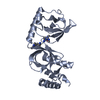
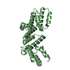
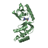
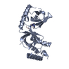
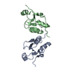
| Entry | Database: PDB / ID: 4k0u | |||||||||
|---|---|---|---|---|---|---|---|---|---|---|
| Title | Pilotin/secretin peptide Complex | |||||||||
 Components Components |
| |||||||||
 Keywords Keywords |  PROTEIN TRANSPORT / Type II secretion system / pilotin-secretin complex / Outer membrane / Dickeya dadantii PROTEIN TRANSPORT / Type II secretion system / pilotin-secretin complex / Outer membrane / Dickeya dadantii | |||||||||
| Function / homology |  Function and homology informationprotein secretion by the type II secretion system / type II protein secretion system complex / cell outer membrane / intracellular protein transport / protein processing / membrane => GO:0016020 Function and homology informationprotein secretion by the type II secretion system / type II protein secretion system complex / cell outer membrane / intracellular protein transport / protein processing / membrane => GO:0016020Similarity search - Function | |||||||||
| Biological species |  Dickeya dadantii (bacteria) Dickeya dadantii (bacteria) | |||||||||
| Method | X-RAY DIFFRACTION / SYNCHROTRON / MOLECULAR REPLACEMENT / Resolution: 2.15 Å | |||||||||
 Authors Authors | Rehman, S. / Pickersgill, R.W. | |||||||||
 Citation Citation | Journal: Acta Crystallogr.,Sect.D / Year: 2013 Title: Anatomy of secretin binding to the Dickeya dadantii type II secretion system pilotin. Authors: Rehman, S. / Gu, S. / Shevchik, V.E. / Pickersgill, R.W. #1: Journal: Plos Pathog. / Year: 2012 Title: Structural and functional insights into the pilotin-secretin complex of the type II secretion system. Authors: Gu, S. / Rehman, S. / Wang, X. / Shevchik, V.E. / Pickersgill, R.W. | |||||||||
| History |
|
- Structure visualization
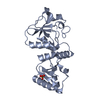
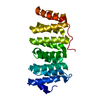
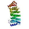
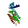
| Structure viewer | Molecule: MolmilJmol/JSmol |
|---|
- Downloads & links
-Download
| PDBx/mmCIF format | 4k0u.cif.gz | 35 KB | Display | PDBx/mmCIF format |
|---|---|---|---|---|
| PDB format | pdb4k0u.ent.gz | 23.4 KB | Display | PDB format |
| PDBx/mmJSON format | 4k0u.json.gz | Tree view | PDBx/mmJSON format | |
| Others |  Other downloads Other downloads |
-Validation report
| Arichive directory | https://data.pdbj.org/pub/pdb/validation_reports/k0/4k0uftp://data.pdbj.org/pub/pdb/validation_reports/k0/4k0u | HTTPS FTP |
|---|
-Related structure data
| Related structure data |  3utkS S: Starting model for refinement |
|---|---|
| Similar structure data |

-Links
 PDBj
PDBj


- Assembly
| Deposited unit | 
| ||||||||
|---|---|---|---|---|---|---|---|---|---|
| 1 | 
| ||||||||
| 2 |
| ||||||||
| Unit cell |
| ||||||||
| Components on special symmetry positions |
|
-Components
| #1: Protein | Mass: 11591.064 Da / Num. of mol.: 1 / Fragment: UNP residues 28-133 Source method: isolated from a genetically manipulated source Source: (gene. exp.) Dickeya dadantii (bacteria) / Strain: 3937 / Gene: Dda3937_02411, outS, OutS Dda3937_02411 / Production host: Escherichia coli (E. coli) / References: UniProt: Q01567 |
|---|---|
| #2: Protein/peptide | Type II secretion system / T2SS protein D / General secretion pathway protein D / Pectic enzymes secretion protein OutD Mass: 1850.983 Da / Num. of mol.: 1 / Fragment: UNP residues 693-705 / Source method: obtained synthetically Details: Synthetic peptide C-terminal OutD (Dickeya dadantii 3937) Source: (synth.) Dickeya dadantii (bacteria) / References: UniProt: Q01565 |
| #3: Water | ChemComp-HOH / Water Mass: 18.015 Da / Num. of mol.: 14 / Source method: isolated from a natural source / Formula: H2O Mass: 18.015 Da / Num. of mol.: 14 / Source method: isolated from a natural source / Formula: H2O |
| Sequence details | RESIDUES ARG B 1 AND ASP B 15 WERE ADDED TO THE SYNTHETIC PEPTIDE TO AID SOLUBILITY |
-Experimental details
-Experiment
| Experiment | Method: X-RAY DIFFRACTION / Number of used crystals: 1 |
|---|
- Sample preparation
| Crystal | Density Matthews: 2.2 Å3/Da / Density % sol: 44.02 % |
|---|---|
| Crystal grow | Temperature: 293 K / Method: vapor diffusion, sitting drop / pH: 7.5 Details: 2% PEG 400, 0.1M HEPES sodium, 2M ammonium sulphate, pH 7.5, VAPOR DIFFUSION, SITTING DROP, temperature 293K |
-Data collection
| Diffraction | Mean temperature: 100 K |
|---|---|
| Diffraction source | Source: SYNCHROTRON / Site: SOLEIL  / Beamline: PROXIMA 1 / Wavelength: 1.0718 Å / Beamline: PROXIMA 1 / Wavelength: 1.0718 Å |
| Detector | Type: DECTRIS PILATUS 6M / Detector: PIXEL / Date: Feb 16, 2012 |
| Radiation | Monochromator: Si 111 CHANNEL / Protocol: SINGLE WAVELENGTH / Monochromatic (M) / Laue (L): M / Scattering type: x-ray |
| Radiation wavelength | Wavelength: 1.0718 Å / Relative weight: 1 |
| Reflection | Resolution: 2.15→47.48 Å / Num. obs: 7209 / % possible obs: 100 % / Observed criterion σ(F): 0 / Observed criterion σ(I): 0 / Redundancy: 7.9 % / Biso Wilson estimate: 27.5 Å2 / Rmerge(I) obs: 0.137 / Rsym value: 0.137 / Net I/σ(I): 9.1 |
| Reflection shell | Resolution: 2.15→2.27 Å / Redundancy: 8.1 % / Rmerge(I) obs: 0.804 / Mean I/σ(I) obs: 2.4 / Num. unique all: 1009 / Rsym value: 0.84 / % possible all: 100 |
- Processing
| Software |
| ||||||||||||||||||||||||||||||||||||||||||||||||||||||||||||||||||||||||||||||||||||||||||||||||||||||||||||||||||||||||||||||||||||||||||||||||||||||||||||||||||||||||||
|---|---|---|---|---|---|---|---|---|---|---|---|---|---|---|---|---|---|---|---|---|---|---|---|---|---|---|---|---|---|---|---|---|---|---|---|---|---|---|---|---|---|---|---|---|---|---|---|---|---|---|---|---|---|---|---|---|---|---|---|---|---|---|---|---|---|---|---|---|---|---|---|---|---|---|---|---|---|---|---|---|---|---|---|---|---|---|---|---|---|---|---|---|---|---|---|---|---|---|---|---|---|---|---|---|---|---|---|---|---|---|---|---|---|---|---|---|---|---|---|---|---|---|---|---|---|---|---|---|---|---|---|---|---|---|---|---|---|---|---|---|---|---|---|---|---|---|---|---|---|---|---|---|---|---|---|---|---|---|---|---|---|---|---|---|---|---|---|---|---|---|---|
| Refinement | Method to determine structure: MOLECULAR REPLACEMENT Starting model: PDB ENTRY 3UTK Resolution: 2.15→46.42 Å / Cor.coef. Fo:Fc: 0.956 / Cor.coef. Fo:Fc free: 0.918 / SU B: 7.019 / SU ML: 0.173 / Cross valid method: THROUGHOUT / σ(F): 0 / σ(I): 0 / ESU R: 0.237 / ESU R Free: 0.217 / Stereochemistry target values: MAXIMUM LIKELIHOOD / Details: HYDROGENS HAVE BEEN ADDED IN THE RIDING POSITIONS
| ||||||||||||||||||||||||||||||||||||||||||||||||||||||||||||||||||||||||||||||||||||||||||||||||||||||||||||||||||||||||||||||||||||||||||||||||||||||||||||||||||||||||||
| Solvent computation | Ion probe radii: 0.8 Å / Shrinkage radii: 0.8 Å / VDW probe radii: 1.2 Å / Solvent model: MASK | ||||||||||||||||||||||||||||||||||||||||||||||||||||||||||||||||||||||||||||||||||||||||||||||||||||||||||||||||||||||||||||||||||||||||||||||||||||||||||||||||||||||||||
| Displacement parameters | Biso mean: 51.629 Å2
| ||||||||||||||||||||||||||||||||||||||||||||||||||||||||||||||||||||||||||||||||||||||||||||||||||||||||||||||||||||||||||||||||||||||||||||||||||||||||||||||||||||||||||
| Refinement step | Cycle: LAST / Resolution: 2.15→46.42 Å
| ||||||||||||||||||||||||||||||||||||||||||||||||||||||||||||||||||||||||||||||||||||||||||||||||||||||||||||||||||||||||||||||||||||||||||||||||||||||||||||||||||||||||||
| Refine LS restraints |
| ||||||||||||||||||||||||||||||||||||||||||||||||||||||||||||||||||||||||||||||||||||||||||||||||||||||||||||||||||||||||||||||||||||||||||||||||||||||||||||||||||||||||||
| LS refinement shell | Resolution: 2.15→2.206 Å / Total num. of bins used: 20
|
