 Movie
Movie Controller


+ Open data

- Basic information
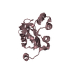
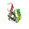
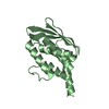
| Entry | Database: PDB / ID: 4k08 | ||||||
|---|---|---|---|---|---|---|---|
| Title | Periplasmic sensor domain of chemotaxis protein, Adeh_3718 | ||||||
 Components Components | Methyl-accepting chemotaxis sensory transducer | ||||||
 Keywords Keywords |  SIGNALING PROTEIN / methyl accepting chemotaxis / Anaeromyxobacter dehalogenans / PAS-like sensor domain / Structural Genomics / PSI-Biology / Midwest Center for Structural Genomics / MCSG / periplasmic sensor domain SIGNALING PROTEIN / methyl accepting chemotaxis / Anaeromyxobacter dehalogenans / PAS-like sensor domain / Structural Genomics / PSI-Biology / Midwest Center for Structural Genomics / MCSG / periplasmic sensor domain | ||||||
| Function / homology |  Function and homology information Function and homology informationmembrane => GO:0016020 / signal transduction / metal ion binding / plasma membraneSimilarity search - Function | ||||||
| Biological species |  Anaeromyxobacter dehalogenans (bacteria) Anaeromyxobacter dehalogenans (bacteria) | ||||||
| Method | X-RAY DIFFRACTION / SYNCHROTRON / SAD / Resolution: 2 Å | ||||||
 Authors Authors | Pokkuluri, P.R. / Mack, J.C. / Bearden, J. / Rakowski, E. / Schiffer, M. / Joachimiak, A. / Midwest Center for Structural Genomics (MCSG) | ||||||
 Citation Citation | Journal: Microbiologyopen / Year: 2013 Title: Analysis of periplasmic sensor domains from Anaeromyxobacter dehalogenans 2CP-C: structure of one sensor domain from a histidine kinase and another from a chemotaxis protein. Authors: Pokkuluri, P.R. / Dwulit-Smith, J. / Duke, N.E. / Wilton, R. / Mack, J.C. / Bearden, J. / Rakowski, E. / Babnigg, G. / Szurmant, H. / Joachimiak, A. / Schiffer, M. | ||||||
| History |
|
- Structure visualization
| Structure viewer | Molecule: MolmilJmol/JSmol |
|---|
- Downloads & links
-Download
| PDBx/mmCIF format | 4k08.cif.gz | 70.4 KB | Display | PDBx/mmCIF format |
|---|---|---|---|---|
| PDB format | pdb4k08.ent.gz | 56 KB | Display | PDB format |
| PDBx/mmJSON format | 4k08.json.gz | Tree view | PDBx/mmJSON format | |
| Others |  Other downloads Other downloads |
-Validation report
| Arichive directory | https://data.pdbj.org/pub/pdb/validation_reports/k0/4k08ftp://data.pdbj.org/pub/pdb/validation_reports/k0/4k08 | HTTPS FTP |
|---|
-Related structure data


-Links
 PDBj
PDBj

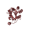
- Assembly
| Deposited unit | 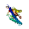
| ||||||||
|---|---|---|---|---|---|---|---|---|---|
| 1 |
| ||||||||
| 2 | 
| ||||||||
| Unit cell |
|
-Components
| #1: Protein | Mass: 17317.023 Da / Num. of mol.: 1 / Fragment: Periplasmic sensor domain (UNP residues 38-190) Source method: isolated from a genetically manipulated source Source: (gene. exp.) Anaeromyxobacter dehalogenans (bacteria)Strain: 2CP-C / Gene: Adeh_3718 / Production host: Escherichia coli (E. coli) / References: UniProt: Q2IFX2 |
|---|---|
| #2: Chemical | ChemComp-ZN /   Mass: 65.409 Da / Num. of mol.: 1 / Source method: obtained synthetically / Formula: Zn Mass: 65.409 Da / Num. of mol.: 1 / Source method: obtained synthetically / Formula: Zn |
| #3: Chemical | ChemComp-ACT / Acetate  Mass: 59.044 Da / Num. of mol.: 1 / Source method: obtained synthetically / Formula: C2H3O2 Mass: 59.044 Da / Num. of mol.: 1 / Source method: obtained synthetically / Formula: C2H3O2 |
| #4: Water | ChemComp-HOH / Water Mass: 18.015 Da / Num. of mol.: 76 / Source method: isolated from a natural source / Formula: H2O Mass: 18.015 Da / Num. of mol.: 76 / Source method: isolated from a natural source / Formula: H2O |
-Experimental details
-Experiment
| Experiment | Method: X-RAY DIFFRACTION / Number of used crystals: 1 |
|---|
- Sample preparation
| Crystal | Density Matthews: 2.41 Å3/Da / Density % sol: 48.99 % |
|---|---|
| Crystal grow | Temperature: 298 K / Method: vapor diffusion, hanging drop / pH: 4.6 Details: Membfac-2 (12% PEG4000, 0.1M sodium acetate pH 4.6, 0.1 M zinc acetate) diluted by water; (membfac-2, 350 uL + water, 150 uL), VAPOR DIFFUSION, HANGING DROP, temperature 298K |
-Data collection
| Diffraction | Mean temperature: 100 K |
|---|---|
| Diffraction source | Source: SYNCHROTRON / Site: APS  / Beamline: 19-BM / Wavelength: 0.97911 Å / Beamline: 19-BM / Wavelength: 0.97911 Å |
| Detector | Detector: CCD / Date: Nov 23, 2010 |
| Radiation | Protocol: SINGLE WAVELENGTH / Monochromatic (M) / Laue (L): M / Scattering type: x-ray |
| Radiation wavelength | Wavelength: 0.97911 Å / Relative weight: 1 |
| Reflection | Resolution: 2→100 Å / Num. obs: 21845 / % possible obs: 99.9 % / Observed criterion σ(I): -3 / Redundancy: 5.8 % / Rmerge(I) obs: 0.03 / Net I/σ(I): 53 |
| Reflection shell | Resolution: 2→2.02 Å / Redundancy: 4.6 % / Rmerge(I) obs: 0.196 / Mean I/σ(I) obs: 8 / % possible all: 100 |
- Processing
| Software |
| |||||||||||||||||||||||||||||||||||||||||||||||||||||||||||||||||||||||||||||||||||||||||||||||||||||||||||||||||||||||||||||
|---|---|---|---|---|---|---|---|---|---|---|---|---|---|---|---|---|---|---|---|---|---|---|---|---|---|---|---|---|---|---|---|---|---|---|---|---|---|---|---|---|---|---|---|---|---|---|---|---|---|---|---|---|---|---|---|---|---|---|---|---|---|---|---|---|---|---|---|---|---|---|---|---|---|---|---|---|---|---|---|---|---|---|---|---|---|---|---|---|---|---|---|---|---|---|---|---|---|---|---|---|---|---|---|---|---|---|---|---|---|---|---|---|---|---|---|---|---|---|---|---|---|---|---|---|---|---|
| Refinement | Method to determine structure: SAD / Resolution: 2→34.594 Å / SU ML: 0.53 / σ(F): 1.34 / Phase error: 21.3 / Stereochemistry target values: MLHL / Details: Hydrogens were added in the riding positions
| |||||||||||||||||||||||||||||||||||||||||||||||||||||||||||||||||||||||||||||||||||||||||||||||||||||||||||||||||||||||||||||
| Solvent computation | Shrinkage radii: 0.77 Å / VDW probe radii: 0.9 Å / Solvent model: FLAT BULK SOLVENT MODEL / Bsol: 40.13 Å2 / ksol: 0.4 e/Å3 | |||||||||||||||||||||||||||||||||||||||||||||||||||||||||||||||||||||||||||||||||||||||||||||||||||||||||||||||||||||||||||||
| Displacement parameters |
| |||||||||||||||||||||||||||||||||||||||||||||||||||||||||||||||||||||||||||||||||||||||||||||||||||||||||||||||||||||||||||||
| Refinement step | Cycle: LAST / Resolution: 2→34.594 Å
| |||||||||||||||||||||||||||||||||||||||||||||||||||||||||||||||||||||||||||||||||||||||||||||||||||||||||||||||||||||||||||||
| Refine LS restraints |
| |||||||||||||||||||||||||||||||||||||||||||||||||||||||||||||||||||||||||||||||||||||||||||||||||||||||||||||||||||||||||||||
| LS refinement shell |
| |||||||||||||||||||||||||||||||||||||||||||||||||||||||||||||||||||||||||||||||||||||||||||||||||||||||||||||||||||||||||||||
| Refinement TLS params. | Method: refined / Refine-ID: X-RAY DIFFRACTION
| |||||||||||||||||||||||||||||||||||||||||||||||||||||||||||||||||||||||||||||||||||||||||||||||||||||||||||||||||||||||||||||
| Refinement TLS group |
|
