 Movie
Movie Controller

[English] 日本語
 Yorodumi
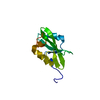
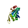
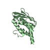
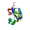
- PDB-2rjn: Crystal structure of an uncharacterized protein Q2BKU2 from Neptu... -
+ Open data

- Basic information
| Entry | Database: PDB / ID: 2rjn | ||||||
|---|---|---|---|---|---|---|---|
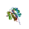
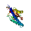
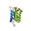
| Title | Crystal structure of an uncharacterized protein Q2BKU2 from Neptuniibacter caesariensis | ||||||
 Components Components | Response regulator receiver:Metal-dependent phosphohydrolase, HD subdomain | ||||||
 Keywords Keywords |  HYDROLASE / STRUCTURAL GENOMICS / Oceanospirillum sp. MED92 / Neptuniibacter caesariensis / PSI-2 / PROTEIN STRUCTURE INITIATIVE / New York SGX Research Center for Structural Genomics / NYSGXRC HYDROLASE / STRUCTURAL GENOMICS / Oceanospirillum sp. MED92 / Neptuniibacter caesariensis / PSI-2 / PROTEIN STRUCTURE INITIATIVE / New York SGX Research Center for Structural Genomics / NYSGXRC | ||||||
| Function / homology | Response regulator / Rossmann fold / 3-Layer(aba) Sandwich / Alpha Beta / Response regulator receiver:Metal-dependent phosphohydrolase, HD subdomain / Response regulator receiver:Metal-dependent phosphohydrolase, HD subdomain Function and homology information Function and homology information | ||||||
| Biological species |  Neptuniibacter caesariensis (bacteria) Neptuniibacter caesariensis (bacteria) | ||||||
| Method | X-RAY DIFFRACTION / SYNCHROTRON / SAD / Resolution: 2.1 Å | ||||||
 Authors Authors | Malashkevich, V.N. / Toro, R. / Meyer, A.J. / Sauder, J.M. / Burley, S.K. / Almo, S.C. / New York SGX Research Center for Structural Genomics (NYSGXRC) | ||||||
 Citation Citation | Journal: To be Published Title: Crystal structure of an uncharacterized protein Q2BKU2 from Neptuniibacter caesariensis. Authors: Malashkevich, V.N. / Toro, R. / Meyer, A.J. / Sauder, J.M. / Burley, S.K. / Almo, S.C. | ||||||
| History |
|
- Structure visualization
| Structure viewer | Molecule: MolmilJmol/JSmol |
|---|
- Downloads & links
-Download
| PDBx/mmCIF format | 2rjn.cif.gz | 42.2 KB | Display | PDBx/mmCIF format |
|---|---|---|---|---|
| PDB format | pdb2rjn.ent.gz | 28.8 KB | Display | PDB format |
| PDBx/mmJSON format | 2rjn.json.gz | Tree view | PDBx/mmJSON format | |
| Others |  Other downloads Other downloads |
-Validation report
| Arichive directory | https://data.pdbj.org/pub/pdb/validation_reports/rj/2rjnftp://data.pdbj.org/pub/pdb/validation_reports/rj/2rjn | HTTPS FTP |
|---|
-Related structure data
| Similar structure data | |
|---|---|
| Other databases |


-Links
 PDBj
PDBj- Assembly
| Deposited unit | 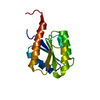
| ||||||||
|---|---|---|---|---|---|---|---|---|---|
| 1 |
| ||||||||
| Unit cell |
| ||||||||
| Components on special symmetry positions |
| ||||||||
| Details | Authors state that the biological unit is unknown. |
-Components
| #1: Protein | Mass: 17657.207 Da / Num. of mol.: 1 / Fragment: Residues 2-144 Source method: isolated from a genetically manipulated source Source: (gene. exp.) Neptuniibacter caesariensis (bacteria) / Strain: MED92 / Gene: MED92_01309 / Plasmid: BC-pSGX3(BC) / Production host: Escherichia coli (E. coli) / Strain (production host): BL21(DE3)Codon+RIL / References: UniProt: Q2BKU2, UniProt: A0A7U8C3V7*PLUS |
|---|---|
| #2: Water | ChemComp-HOH / Water Mass: 18.015 Da / Num. of mol.: 64 / Source method: isolated from a natural source / Formula: H2O Mass: 18.015 Da / Num. of mol.: 64 / Source method: isolated from a natural source / Formula: H2O |
-Experimental details
-Experiment
| Experiment | Method: X-RAY DIFFRACTION / Number of used crystals: 1 |
|---|
- Sample preparation
| Crystal | Density Matthews: 2.69 Å3/Da / Density % sol: 54.32 % |
|---|---|
| Crystal grow | Temperature: 294 K / Method: vapor diffusion, sitting drop Details: 4 M Sodium formate, 0.01M Spermine tetrachloride, VAPOR DIFFUSION, SITTING DROP, temperature 294K |
-Data collection
| Diffraction | Mean temperature: 100 K | |||||||||||||||||||||||||||||||||||||||||||||||||||||||||||||||||||||||||||||
|---|---|---|---|---|---|---|---|---|---|---|---|---|---|---|---|---|---|---|---|---|---|---|---|---|---|---|---|---|---|---|---|---|---|---|---|---|---|---|---|---|---|---|---|---|---|---|---|---|---|---|---|---|---|---|---|---|---|---|---|---|---|---|---|---|---|---|---|---|---|---|---|---|---|---|---|---|---|---|
| Diffraction source | Source: SYNCHROTRON / Site: NSLS  / Beamline: X29A / Wavelength: 0.979 Å / Beamline: X29A / Wavelength: 0.979 Å | |||||||||||||||||||||||||||||||||||||||||||||||||||||||||||||||||||||||||||||
| Detector | Type: ADSC QUANTUM 315 / Detector: CCD / Date: Oct 11, 2007 / Details: X29 | |||||||||||||||||||||||||||||||||||||||||||||||||||||||||||||||||||||||||||||
| Radiation | Protocol: SINGLE WAVELENGTH / Monochromatic (M) / Laue (L): M / Scattering type: x-ray | |||||||||||||||||||||||||||||||||||||||||||||||||||||||||||||||||||||||||||||
| Radiation wavelength | Wavelength: 0.979 Å / Relative weight: 1 | |||||||||||||||||||||||||||||||||||||||||||||||||||||||||||||||||||||||||||||
| Reflection | Redundancy: 3.5 % / Av σ(I) over netI: 14.6 / Number: 68890 / Rmerge(I) obs: 0.059 / Χ2: 2.37 / D res high: 2 Å / D res low: 50 Å / Num. obs: 19935 / % possible obs: 79.9 | |||||||||||||||||||||||||||||||||||||||||||||||||||||||||||||||||||||||||||||
| Diffraction reflection shell |
| |||||||||||||||||||||||||||||||||||||||||||||||||||||||||||||||||||||||||||||
| Reflection | Resolution: 2→50 Å / Num. obs: 19935 / % possible obs: 79.9 % / Redundancy: 3.5 % / Rmerge(I) obs: 0.059 / Χ2: 2.372 / Net I/σ(I): 14.6 | |||||||||||||||||||||||||||||||||||||||||||||||||||||||||||||||||||||||||||||
| Reflection shell |
|
-Phasing
| Phasing | Method: SAD |
|---|
- Processing
| Software |
| ||||||||||||||||||||||||||||||||||||||||||||||||||||||||||||||||||||||||||||||||||||||||||
|---|---|---|---|---|---|---|---|---|---|---|---|---|---|---|---|---|---|---|---|---|---|---|---|---|---|---|---|---|---|---|---|---|---|---|---|---|---|---|---|---|---|---|---|---|---|---|---|---|---|---|---|---|---|---|---|---|---|---|---|---|---|---|---|---|---|---|---|---|---|---|---|---|---|---|---|---|---|---|---|---|---|---|---|---|---|---|---|---|---|---|---|
| Refinement | Method to determine structure: SAD / Resolution: 2.1→19.23 Å / Cor.coef. Fo:Fc: 0.956 / Cor.coef. Fo:Fc free: 0.921 / SU B: 9.36 / SU ML: 0.116 / TLS residual ADP flag: LIKELY RESIDUAL / Cross valid method: THROUGHOUT / σ(F): 0 / ESU R: 0.197 / ESU R Free: 0.178 / Stereochemistry target values: MAXIMUM LIKELIHOOD Details: The Bijvoet differences were used in phasing. HYDROGENS HAVE BEEN ADDED IN THE RIDING POSITIONS
| ||||||||||||||||||||||||||||||||||||||||||||||||||||||||||||||||||||||||||||||||||||||||||
| Solvent computation | Ion probe radii: 0.8 Å / Shrinkage radii: 0.8 Å / VDW probe radii: 1.2 Å / Solvent model: BABINET MODEL WITH MASK | ||||||||||||||||||||||||||||||||||||||||||||||||||||||||||||||||||||||||||||||||||||||||||
| Displacement parameters | Biso mean: 39.612 Å2
| ||||||||||||||||||||||||||||||||||||||||||||||||||||||||||||||||||||||||||||||||||||||||||
| Refinement step | Cycle: LAST / Resolution: 2.1→19.23 Å
| ||||||||||||||||||||||||||||||||||||||||||||||||||||||||||||||||||||||||||||||||||||||||||
| Refine LS restraints |
| ||||||||||||||||||||||||||||||||||||||||||||||||||||||||||||||||||||||||||||||||||||||||||
| LS refinement shell | Resolution: 2.1→2.154 Å / Total num. of bins used: 20
| ||||||||||||||||||||||||||||||||||||||||||||||||||||||||||||||||||||||||||||||||||||||||||
| Refinement TLS params. | Method: refined / Origin x: -0.8908 Å / Origin y: 21.1673 Å / Origin z: 31.998 Å
|
