Mass: 18.015 Da / Num. of mol.: 348 / Source method: isolated from a natural source / Formula: H2O
-
Experimental details
-
Experiment
Experiment
Method: X-RAY DIFFRACTION / Number of used crystals: 1
-
Sample preparation
Crystal
Density Matthews: 2.16 Å3/Da / Density % sol: 43.12 %
Crystal grow
Temperature: 289 K / Method: vapor diffusion, sitting drop Details: The drop was 200 nl 13.75 mg/ml protein mixed with 200 nl Qiagen Cryos Suite condition A4 (4.25 % isopropanol, 1.7 M Ammonium Sulfate, 15% glycerol). The crystallization reservoir contained ...Details: The drop was 200 nl 13.75 mg/ml protein mixed with 200 nl Qiagen Cryos Suite condition A4 (4.25 % isopropanol, 1.7 M Ammonium Sulfate, 15% glycerol). The crystallization reservoir contained only 1.5 M NaCl, VAPOR DIFFUSION, SITTING DROP, temperature 289K
Type: MARMOSAIC 225 mm CCD / Detector: CCD / Date: Mar 9, 2013
Radiation
Monochromator: Diamond [111] / Protocol: SINGLE WAVELENGTH / Monochromatic (M) / Laue (L): M / Scattering type: x-ray
Radiation wavelength
Wavelength: 0.97872 Å / Relative weight: 1
Reflection
Redundancy: 11.1 % / Av σ(I) over netI: 35.66 / Number: 458154 / Rmerge(I) obs: 0.098 / Χ2: 0.92 / D res high: 1.9 Å / D res low: 40 Å / Num. obs: 41251 / % possible obs: 99.5
Diffraction reflection shell
Highest resolution (Å)
Lowest resolution (Å)
% possible obs (%)
ID
Rmerge(I) obs
Chi squared
Redundancy
5.15
40
99.8
1
0.032
0.544
11.6
4.09
5.15
100
1
0.065
1.351
12.3
3.58
4.09
100
1
0.071
1.421
12.4
3.25
3.58
100
1
0.065
0.989
12.4
3.02
3.25
100
1
0.073
0.896
12.5
2.84
3.02
100
1
0.095
0.919
12.5
2.7
2.84
100
1
0.121
0.901
12.5
2.58
2.7
100
1
0.151
0.894
12.5
2.48
2.58
100
1
0.196
0.901
12.5
2.39
2.48
100
1
0.235
0.879
12.5
2.32
2.39
100
1
0.278
0.89
12.5
2.25
2.32
100
1
0.359
0.867
12.5
2.19
2.25
100
1
0.435
0.861
12.5
2.14
2.19
100
1
0.548
0.839
12.4
2.09
2.14
100
1
0.642
0.858
12.3
2.05
2.09
100
1
0.791
0.868
10.1
2.01
2.05
97.8
1
0.881
0.815
8.3
1.97
2.01
97.4
1
0.792
6.9
1.93
1.97
97.6
1
0.795
5.7
1.9
1.93
97.7
1
0.828
4.8
Reflection
Resolution: 2.1→40 Å / Num. obs: 30946 / % possible obs: 100 % / Redundancy: 12.2 % / Rmerge(I) obs: 0.116 / Χ2: 1.721 / Net I/σ(I): 8.2
Reflection shell
Resolution (Å)
Redundancy (%)
Rmerge(I) obs
Num. unique all
Χ2
Diffraction-ID
% possible all
2.1-2.14
12
0.773
1525
0.501
1
100
2.14-2.18
12.2
0.713
1518
0.509
1
100
2.18-2.22
12.4
0.634
1517
0.516
1
100
2.22-2.26
12.4
0.538
1548
0.55
1
100
2.26-2.31
12.5
0.458
1531
0.573
1
100
2.31-2.37
12.5
0.382
1521
0.605
1
100
2.37-2.42
12.5
0.336
1566
0.65
1
100
2.42-2.49
12.5
0.288
1517
0.696
1
100
2.49-2.56
12.5
0.25
1512
0.732
1
100
2.56-2.65
12.5
0.207
1556
0.827
1
100
2.65-2.74
12.6
0.179
1541
0.934
1
100
2.74-2.85
12.5
0.15
1532
1.112
1
100
2.85-2.98
12.5
0.133
1551
1.38
1
100
2.98-3.14
12.5
0.105
1557
1.618
1
100
3.14-3.33
12.4
0.096
1532
2.093
1
100
3.33-3.59
12.3
0.085
1541
2.752
1
100
3.59-3.95
12
0.082
1566
3.803
1
100
3.95-4.52
11.6
0.075
1564
4.372
1
100
4.52-5.7
11.7
0.072
1594
4.758
1
100
5.7-40
11.1
0.067
1657
5.887
1
100
-
Processing
Software
Name
Version
Classification
NB
DENZO
datareduction
SCALEPACK
datascaling
REFMAC
refinement
PDB_EXTRACT
3.11
dataextraction
HKL-3000
datareduction
SHELXS
phasing
Refinement
Method to determine structure: SAD / Resolution: 2.1→38.8 Å / Cor.coef. Fo:Fc: 0.972 / Cor.coef. Fo:Fc free: 0.96 / WRfactor Rfree: 0.1731 / WRfactor Rwork: 0.1394 / Occupancy max: 1 / Occupancy min: 0.33 / FOM work R set: 0.9125 / SU B: 6.302 / SU ML: 0.088 / SU R Cruickshank DPI: 0.1711 / SU Rfree: 0.1437 / Cross valid method: THROUGHOUT / σ(F): 0 / ESU R: 0.171 / ESU R Free: 0.144 / Stereochemistry target values: MAXIMUM LIKELIHOOD Details: U VALUES : WITH TLS ADDED HYDROGENS HAVE BEEN ADDED IN THE RIDING POSITIONS
Rfactor
Num. reflection
% reflection
Selection details
Rfree
0.1793
1552
5 %
RANDOM
Rwork
0.141
-
-
-
obs
0.1429
30862
99.98 %
-
Solvent computation
Ion probe radii: 0.8 Å / Shrinkage radii: 0.8 Å / VDW probe radii: 1.2 Å / Solvent model: MASK
In the structure databanks used in Yorodumi, some data are registered as the other names, "COVID-19 virus" and "2019-nCoV". Here are the details of the virus and the list of structure data.
Jan 31, 2019. EMDB accession codes are about to change! (news from PDBe EMDB page)
EMDB accession codes are about to change! (news from PDBe EMDB page)
The allocation of 4 digits for EMDB accession codes will soon come to an end. Whilst these codes will remain in use, new EMDB accession codes will include an additional digit and will expand incrementally as the available range of codes is exhausted. The current 4-digit format prefixed with “EMD-” (i.e. EMD-XXXX) will advance to a 5-digit format (i.e. EMD-XXXXX), and so on. It is currently estimated that the 4-digit codes will be depleted around Spring 2019, at which point the 5-digit format will come into force.
The EM Navigator/Yorodumi systems omit the EMD- prefix.
Related info.:Q: What is EMD? / ID/Accession-code notation in Yorodumi/EM Navigator
Yorodumi is a browser for structure data from EMDB, PDB, SASBDB, etc.
This page is also the successor to EM Navigator detail page, and also detail information page/front-end page for Omokage search.
The word "yorodu" (or yorozu) is an old Japanese word meaning "ten thousand". "mi" (miru) is to see.
Related info.:EMDB / PDB / SASBDB / Comparison of 3 databanks / Yorodumi Search / Aug 31, 2016. New EM Navigator & Yorodumi / Yorodumi Papers / Jmol/JSmol / Function and homology information / Changes in new EM Navigator and Yorodumi
 Movie
Movie Controller

 Yorodumi
 Open data
 Basic information
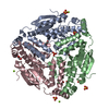
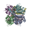
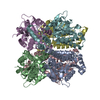
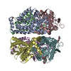
 Components
 Keywords
 LYASE /
  Function and homology information
 Authors
 Citation
 Structure visualization
 Downloads & links
 Other downloads
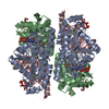
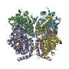
 PDBj
PDBj Assembly

 Mass: 18.015 Da / Num. of mol.: 348 / Source method: isolated from a natural source / Formula: H2O
 Sample preparation
 / Beamline: 21-ID-F / Wavelength: 0.97872 Å
 Processing